

The Polyomaviridae: Contributions of virus structure to our understanding of virus receptors and infectious entry
- PMID: 19157478
- PMCID: PMC2663363
- DOI: 10.1016/j.virol.2008.12.021
The Polyomaviridae: Contributions of virus structure to our understanding of virus receptors and infectious entry
Abstract
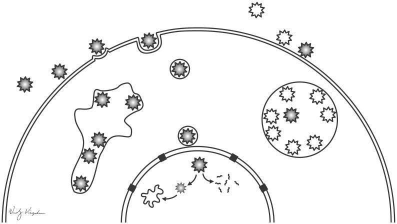
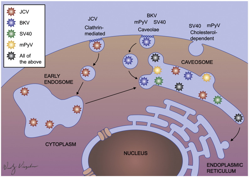
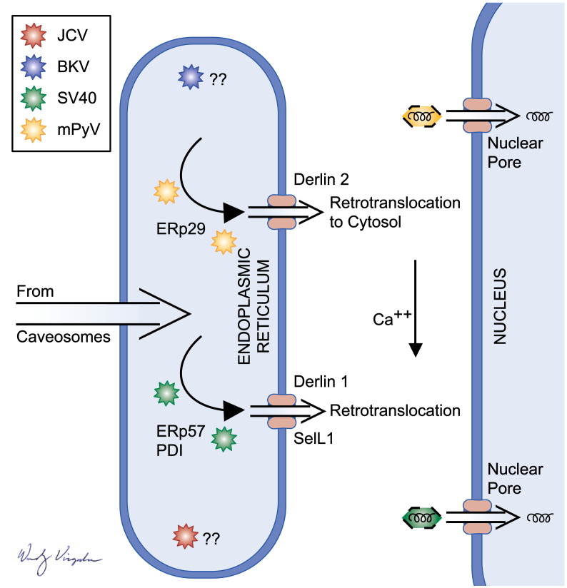
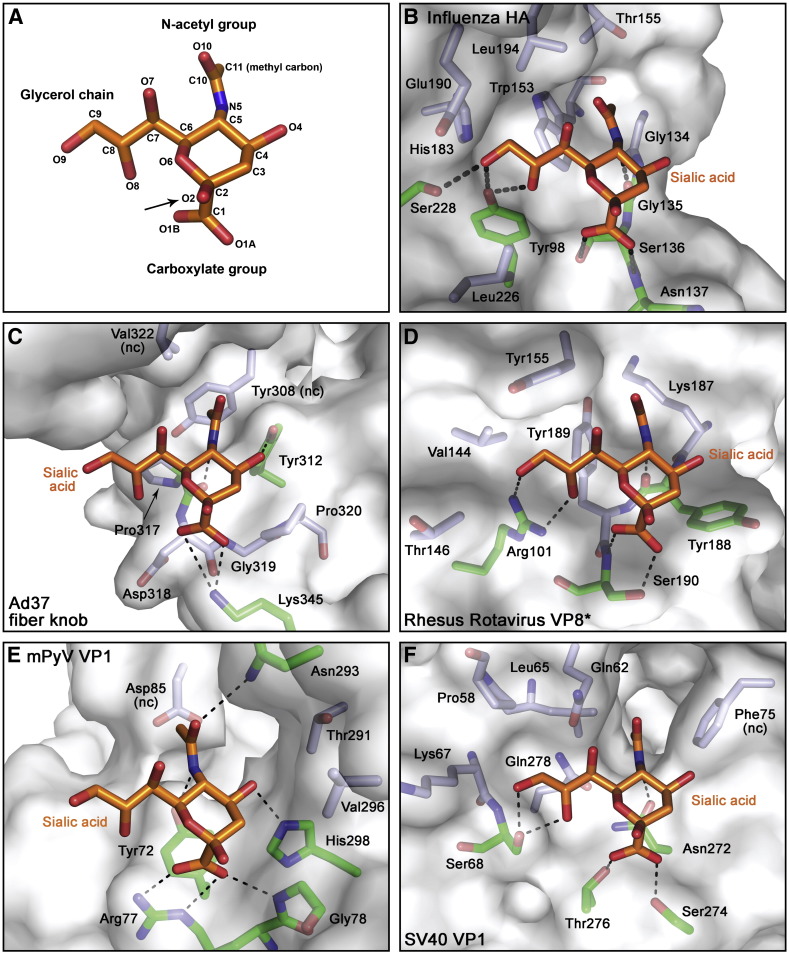
This review summarizes the field's major findings related to the characterization of polyomavirus structures and to the characterization of virus receptors and mechanisms of host cell invasion. The four members of the family that have received the most attention in this regard are the mouse polyomavirus (mPyV), the monkey polyomavirus SV40, and the two human polyomaviruses, JCV and BKV. The structures of both the mPyV and SV40 alone and in complex with receptor fragments have been solved to high resolution. The majority of polyomaviruses recognize terminal sialic acid in either an alpha2,3 linkage or an alpha2,6 linkage to the underlying galactose. Studies on virus structure, receptor utilization and mechanisms of entry have led to new insights into how these viruses interact in an active way with cells to ensure the nuclear delivery and expression of their genomes. Critical work on virus entry has led to the discovery of a pH neutral endocytic compartment that accepts cargo from caveolae and to novel roles for endoplasmic reticulum (ER) associated factors in virus uncoating and penetration of ER membranes. This review will summarize the major findings and compare and contrast the mechanisms used by these viruses to infect cells.
Figures
Similar articles
-
Structure analysis of the major capsid proteins of human polyomaviruses 6 and 7 reveals an obstructed sialic acid binding site.J Virol. 2014 Sep;88(18):10831-9. doi: 10.1128/JVI.01084-14. Epub 2014 Jul 9. J Virol. 2014. PMID: 25008942 Free PMC article.
-
Structural Basis and Evolution of Glycan Receptor Specificities within the Polyomavirus Family.mBio. 2020 Jul 28;11(4):e00745-20. doi: 10.1128/mBio.00745-20. mBio. 2020. PMID: 32723915 Free PMC article.
-
The role of sialic acid in human polyomavirus infections.Glycoconj J. 2006 Feb;23(1-2):19-26. doi: 10.1007/s10719-006-5434-z. Glycoconj J. 2006. PMID: 16575519 Review.
-
Taking the Scenic Route: Polyomaviruses Utilize Multiple Pathways to Reach the Same Destination.Viruses. 2020 Oct 15;12(10):1168. doi: 10.3390/v12101168. Viruses. 2020. PMID: 33076363 Free PMC article. Review.
-
Lipids and proteins act in opposing manners to regulate polyomavirus infection.J Virol. 2010 Oct;84(19):9840-52. doi: 10.1128/JVI.01093-10. Epub 2010 Jul 28. J Virol. 2010. PMID: 20668088 Free PMC article.
Cited by
-
Human polyoma viruses and disease with emphasis on clinical BK and JC.J Clin Virol. 2010 Apr;47(4):306-12. doi: 10.1016/j.jcv.2009.12.006. Epub 2010 Jan 8. J Clin Virol. 2010. PMID: 20060360 Free PMC article. Review.
-
The emerging role and significance of circular RNAs in viral infections and antiviral immune responses: possible implication as theranostic agents.RNA Biol. 2021 Jan;18(1):1-15. doi: 10.1080/15476286.2020.1790198. Epub 2020 Jul 13. RNA Biol. 2021. PMID: 32615049 Free PMC article. Review.
-
Principles of polyoma- and papillomavirus uncoating.Med Microbiol Immunol. 2012 Nov;201(4):427-36. doi: 10.1007/s00430-012-0262-1. Epub 2012 Sep 23. Med Microbiol Immunol. 2012. PMID: 23001401 Review.
-
Coinfections of Novel Polyomavirus, Anelloviruses and a Recombinant Strain of Myxoma Virus-MYXV-Tol Identified in Iberian Hares.Viruses. 2020 Mar 20;12(3):340. doi: 10.3390/v12030340. Viruses. 2020. PMID: 32244962 Free PMC article.
-
Sialic Acid Receptors of Viruses.Top Curr Chem. 2015;367:1-28. doi: 10.1007/128_2013_466. Top Curr Chem. 2015. PMID: 23873408 Free PMC article. Review.
References
-
- Blanchard H., Yu X., Coulson B.S., von Itzstein M. Insight into host cell carbohydrate-recognition by human and porcine rotavirus from crystal structures of the virion spike associated carbohydrate-binding domain (VP8⁎) J. Mol. Biol. 2007;367(4):1215–1226. - PubMed
Further reading
-
- Please see the manuscript entitled “Structure, attachment and entry of polyoma- and Papillomaviruses” in this issue, which also describes polyoma virus structure and entry.
Publication types
MeSH terms
Substances
Grants and funding
LinkOut - more resources
Full Text Sources
Other Literature Sources
