Review
. 2009 Feb;19(1):14-22.
doi: 10.1016/j.sbi.2008.12.007. Epub 2009 Jan 20.

Prion protein misfolding and disease

Affiliations
Review

Prion protein misfolding and disease

Roger A Moore et al. Curr Opin Struct Biol. 2009 Feb.

Abstract

Transmissible spongiform encephalopathies (TSEs or prion diseases) are a rare group of invariably fatal neurodegenerative disorders that affect humans and other mammals. TSEs are protein misfolding diseases that involve the accumulation of an abnormally aggregated form of the normal host prion protein (PrP). They are unique among protein misfolding disorders in that they are transmissible and have different strains of infectious agents that are associated with unique phenotypes in vivo. A wealth of biological and biophysical evidence now suggests that the molecular basis for prion diseases may be encoded by protein conformation. The purpose of this review is to provide an overview of the existing structural information for PrP within the context of what is known about the biology of prion disease.


Figures

None
Estimated localizations of α-helix and β-sheet secondary structure in PrP-res throughout the amino acid sequence. The numbering (shown at the bottom of the figure) is based upon the human PrP sequence. 1Full-length PrP-sen with predicted secondary structure motifs based upon accumulated NMR structures as discussed in the text. 2Experimental FTIR data obtained with hamster 263K PrP-res and secondary structure estimations performed by the method of (Garnier et al. J. Mol. Biol. 1978). The predicted structure shows an increase in β-sheet character, especially in the region from ~120–190 and at the C-terminus. 3The spiral model, proposed by DeMarco and Daggett, is based upon molecular dynamics modeling of recombinant hamster PrP mutant D147N under low pH conditions. In this model the initial core structure is represented as four parallel and antiparallel β-strands yielding a protofibrillar species containing 34% extended β-sheet like structure. The original α-helices remain largely in their native conformation. 4The β-helix model, obtained by threading the sequence of PrP onto the scaffolds of known proteins with left handed β-helices. This model predicts that individual β-strands arrange into left-handed helices that form an amyloid core spanning residues 89–176. The second and third original α-helices of PrP-sen would remain largely unaffected. The monomers would then assemble into a trimer, forming a repetitive β-helical disc which would in turn polymerize into PrP-res fibrils. 5Human r-PrP90–231 was used to form amyloid-like fibrils. The molecular architecture of the protease resistant core spanning residues 160–220 was derived experimentally based upon site-directed spin labeling coupled with EPR spectroscopy and found to consist of a parallel, in-register β-structure. None of the α-helices from the original PrP-sen retained any native conformation. The β-structure is interspersed with tight turns, which were modeled in to account for the existence of a disulfide bridge and to exclude charged residues from the β-sheet interface.
Figure 1
Figure 1. Critical loop regions of PrP-sen
Key PrP-sen residues in surface-exposed critical loop regions are illustrated in red. Experimental results from different research groups suggest that these key residues may represent contact sites between PrP-sen and PrP-res. A) Ribbon diagram of hamster PrP-sen (pbd 1b10) with residues Met139, Asn155 and Asn170 in red. B) Closer view of residue 139 (residue 138 in mouse PrP), found to be critical for conversion of PrP-sen to PrP-res. C) Expanded view of residue 155, shown in red. Sequence homology between PrP-sen and hamster PrP-res at amino acid residue 155 influenced the conversion efficiency of a protease-resistant product induced by hamster PrP-res [17]. D) Residue 170 is found in the β2 -α2 loop and is important in susceptibility to TSE disease. Figure 1 was created with PyMOL. DeLano, Warren L. The PyMOL Molecular Graphics System (2008) DeLano Scientific, Palo Alto, CA, USA http:/www.pymol.org.
Figure 2
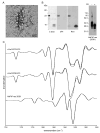
Figure 2. r-HaPrP-res has a different secondary structure than 263K HaPrP-res
Amyloid-like r-PrP-res fibrils made in our laboratory can be PK digested resulting in removal of the N-terminus and generation of a C-terminal protease resistant core. Panel A) shows a representative example of amyloid-like fibrils of hamster r-PrP-sen (residues 23–231) grown at 37 °C with 200rpm in denaturing buffer. B) SDS-PAGE and staining of the PK-treated fibrils with Coomassie blue shows two bands of approximately 8 and 10 kDa, neither of which react with monoclonal antibody 3F4, which is specific for residues 109–112, suggesting that the cleavage site is C-terminal to this epitope. However the 10 kDa band does react with antibody R18, which is specific for residues 142–155, helping to confirm that a 10kDa fragment is present. C) ATR-FTIR spectra of PK-treated r-PrP-res23–231 (from samples shown in panel B) or PrP90–231 show essentially the same β-sheet conformation in the 1610–1640cm−1 region of the spectrum. However, a comparison of these spectra to that of PK-treated PrP-res from hamster TSE strain 263K shows a markedly different banding pattern in this region. A representative immunoblot of PK and non-PK treated 263K PrP-res developed with the monoclonal antibody 3F4 is shown on the far right of panel B.

Similar articles

Cited by

References

    1. Priola SA, Vorberg I. Molecular aspects of disease pathogenesis in the transmissible spongiform encephalopathies. Mol Biotechnol. 2006;33:71–88. - PubMed
    1. Castilla J, Saa P, Hetz C, Soto C. In vitro generation of infectious scrapie prions. Cell. 2005;121:195–206. The first demonstration that infectious prion particles could be generated in vitro by mixing uninfected and prion-infected brain homogenates in serial rounds of dilution and sonication. This method is now widely termed protein misfolding cyclic amplification, or PMCA. - PubMed
    1. Deleault NR, Harris BT, Rees JR, Supattapone S. Formation of native prions from minimal components in vitro. Proc Natl Acad Sci USA. 2007;104:9741–9746. Demonstrated that prions could be formed without infectious PrP-res as a seed from a minimal set of components including native PrP-sen, lipid molecules, and a synthetic polyanion. - PMC - PubMed
    1. Riek R, Hornemann S, Wider G, Billeter M, Glockshuber R, Wuthrich K. NMR structure of the mouse prion protein domain PrP(121–231) Nature. 1996;382:180–182. First NMR solution structure of the globular fold of PrP-sen. - PubMed
    1. Zahn R, Liu A, Luhrs T, Riek R, von Schroetter C, Garcia FL, Billeter M, Calzolai L, Wider G, Wuthrich K. NMR solution structure of human prion protein. Proc Natl Acad Sci USA. 2000;97:145–150. - PMC - PubMed