. 2009 Apr 3;284(14):9299-310.
doi: 10.1074/jbc.M807998200. Epub 2009 Jan 21.

Structure-function relationship of the chloroplastic glutaredoxin S12 with an atypical WCSYS active site

Affiliations

Structure-function relationship of the chloroplastic glutaredoxin S12 with an atypical WCSYS active site

Jeremy Couturier et al. J Biol Chem. .

Abstract

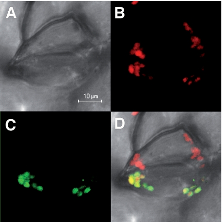
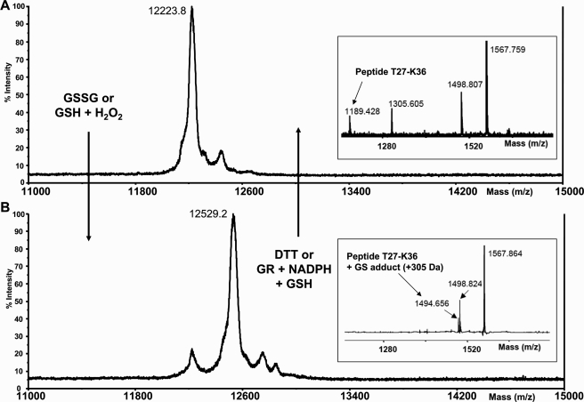
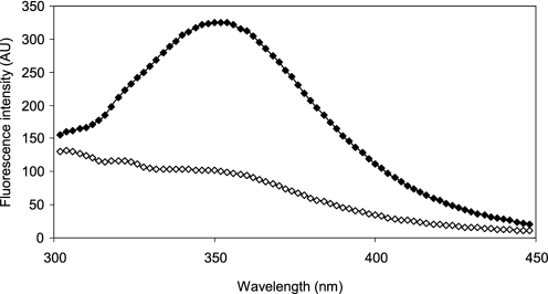
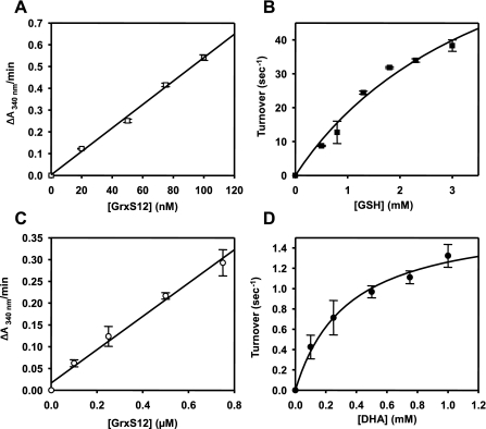
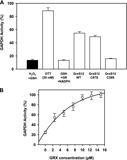
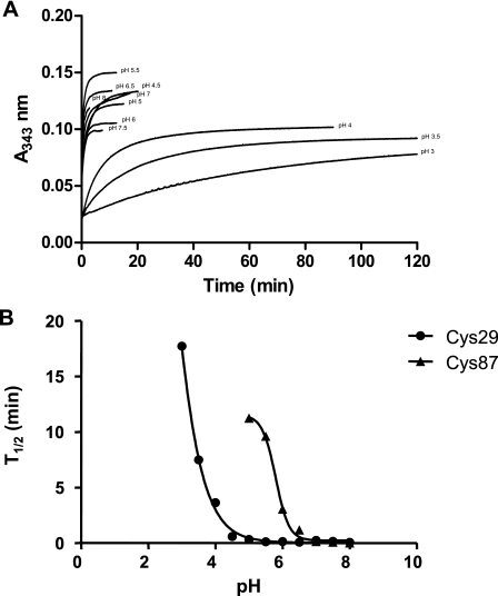
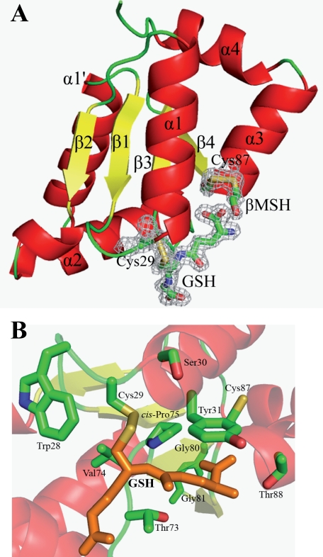
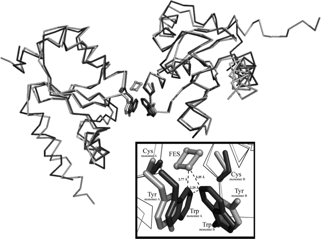
Glutaredoxins (Grxs) are efficient catalysts for the reduction of mixed disulfides in glutathionylated proteins, using glutathione or thioredoxin reductases for their regeneration. Using GFP fusion, we have shown that poplar GrxS12, which possesses a monothiol (28)WCSYS(32) active site, is localized in chloroplasts. In the presence of reduced glutathione, the recombinant protein is able to reduce in vitro substrates, such as hydroxyethyldisulfide and dehydroascorbate, and to regenerate the glutathionylated glyceraldehyde-3-phosphate dehydrogenase. Although the protein possesses two conserved cysteines, it is functioning through a monothiol mechanism, the conserved C terminus cysteine (Cys(87)) being dispensable, since the C87S variant is fully active in all activity assays. Biochemical and crystallographic studies revealed that Cys(87) exhibits a certain reactivity, since its pK(a) is around 5.6. Coupled with thiol titration, fluorescence, and mass spectrometry analyses, the resolution of poplar GrxS12 x-ray crystal structure shows that the only oxidation state is a glutathionylated derivative of the active site cysteine (Cys(29)) and that the enzyme does not form inter- or intramolecular disulfides. Contrary to some plant Grxs, GrxS12 does not incorporate an iron-sulfur cluster in its wild-type form, but when the active site is mutated into YCSYS, it binds a [2Fe-2S] cluster, indicating that the single Trp residue prevents this incorporation.


Figures

FIGURE 1.
FIGURE 1.
GFP localization of GrxS12 in plant guard cells. A, cells under visible light; B, autofluorescence of chlorophyll (red); C, fluorescence of the GFP construction; D, merged images.
FIGURE 2.
FIGURE 2.
MALDI-TOF mass spectrometry analysis of reduced or glutathionylated GrxS12. MALDI-TOF spectra of whole protein or after tryptic digestion (insets) were determined for reduced GrxS12 before (A) or after (B) glutathionylation treatments (5 mm GSSG or 0.1 mm H2O2 plus 0.5 mm GSH for 1 h). The shifted peptide (Thr27–Lys36) is indicated. The 305 Da shift after glutathionylation treatment could be reversed by treatments with 10 mm DTT or with 2 mm GSH in the presence of 6 μg/ml yeast glutathione reductase and 0.5 mm NADPH. Similar results were obtained with the WT and C87S GrxS12.
FIGURE 3.
FIGURE 3.
Fluorescence spectra of poplar GrxS12 under different redox states. Emission spectra of reduced (♦) and oxidized (⋄) GrxS12 (excitation at 290 nm) were recorded with 10 μm protein at 25 °C in TE, pH 8.0, buffer. The reduced GrxS12 was obtained by a pretreatment of the protein with 10 mm DTT. GrxS12 was oxidized using 1 mm GSSG after DTT prereduction. In each case, residual compounds were removed by gel filtration.
FIGURE 4.
FIGURE 4.
HED and DHA reductase activity of poplar GrxS12. A and C, linear dependence of HED (A) and DHA reductase (C) activity on GrxS12 concentration expressed as ΔA340/min. The data are represented as mean ± S.D. B and D, variations of the apparent turnover number during an HED assay catalyzed by 50 nm GrxS12 in the presence of GSH concentrations ranging from 0.5 to 3 mm (B) and during DHA reduction catalyzed by 0.25 μm GrxS12 in the presence of varying DHA concentrations ranging from 0.1 to 1 mm (D). Turnover represents mol of NADPH oxidized/s by 1 mol of GrxS12. Activity was calculated after subtracting the spontaneous reduction rate observed in the absence of GrxS12. Three separate experiments were performed, and the data are represented as mean ± S.D. The best fit was obtained using the Michaelis-Menten equation.
FIGURE 5.
FIGURE 5.
Reactivation of glutathionylated A4-GAPDH. A4-GAPDH was inactivated by incubation with 0.1 mm H2O2 in the presence of 0.5 mm GSH for 15 min at 25 °C and subsequently treated with 35 μm 1,3-bisphosphoglycerate. A, the reactivation assays were performed under the following conditions: (i) 20 mm DTT, (ii) 2 mm GSH in the presence of 6 μg/ml yeast glutathione reductase and 0.2 mm NADPH alone or (iii) in the presence of 10 μm GrxS12 WT, C29S, or C87S. The NADPH-dependent activity was determined before (black bar) and after the different reactivation treatments (white bars). B, reactivation of glutathionylated A4-GAPDH with 2 mm GSH, 6 μg/ml yeast glutathione reductase, 0.2 mm NADPH in the presence of varying concentrations of GrxS12 ranging from 2.5 to 15 μm. Activities are represented as a percentage of the initial activity measured before the inactivation treatment. The data are shown as mean ± S.D.
FIGURE 6.
FIGURE 6.
pKa determination of GrxS12 sulfhydryl groups. A, reaction of GrxS12 C87S with PDT-bimane was monitored at 343 nm at pH values ranging from 3.0 to 8.0. The increase at 343 nm results from the release of pyridyl-2-thione from PDT-bimane. Each curve was fit to the Michaelis-Menten equation. B, t½ for the reactions of PDT-bimane with GrxS12 C29S and C87S were plotted as a function of pH, and the results were fitted to a sigmoidal curve. From this plot, sulfhydryl pKa values of 2.84 ± 0.08 and 5.63 ± 0.17 were determined for Cys29 and Cys87, respectively.
FIGURE 7.
FIGURE 7.
Schematic representations of GrxS12·GSH·βMSH structure (A), highlighting the active site of the protein in B. All α-helices are shown in red, whereas β-strands are in yellow and connecting loops are in green. Corresponding secondary structures are labeled. Both Cys29 and Cys87, GSH, and βMSH molecules are highlighted in sticks, with final 2Fo - Fc electron densities (1.2σ level) covering chosen residues and ligands for clarity. The catalytic Cys29 and the bound GSH molecule are highlighted in orange in B for clarity.
FIGURE 8.
FIGURE 8.
Superimposition of the hypothetical dimer of GrxS12 (dark gray) to the poplar GrxC1 dimer (light gray). A close-up view detailing the ISC at the active site of poplar GrxC1 is presented in the inset. Upon the superimposition of the hypothetical dimer of GrxS12, Trp28 of GrxS12 is the major hindrance of dimerization and the incorporation of the ISC. The dimer of poplar GrxC1 is bridged by a [2Fe-2S] cluster depicted as spheres with sticks (light gray) showing the coordination. The close distances between the ISC and both Trp residues of GrxS12 are labeled.

References

    1. Bushweller, J. H., Aslund, F., Wuthrich, K., and Holmgren, A. (1992) Biochemistry 31 9288-9293 - PubMed
    1. Rouhier, N., Gelhaye, E., and Jacquot, J. P. (2002) FEBS Lett. 511 145-149 - PubMed
    1. Lillig, C. H., Prior, A., Schwenn, J. D., Aslund, F., Ritz, D., Vlamis-Gardikas, A., and Holmgren, A. (1999) J. Biol. Chem. 74 7695-7698 - PubMed
    1. Johansson, C., Lillig, C. H., and Holmgren, A. (2004) J. Biol. Chem. 279 7537-7543 - PubMed
    1. Tamarit, J., Bellí, G., Cabiscol, E., Herrero, E., and Ros, J. (2003) J. Biol. Chem. 278 25745-25751 - PubMed

Publication types

MeSH terms