. 2009 Apr 3;284(14):9529-39.
doi: 10.1074/jbc.M809393200. Epub 2009 Jan 21.

BMP4 mediates oxidative stress-induced retinal pigment epithelial cell senescence and is overexpressed in age-related macular degeneration

Affiliations

BMP4 mediates oxidative stress-induced retinal pigment epithelial cell senescence and is overexpressed in age-related macular degeneration

Danhong Zhu et al. J Biol Chem. .

Abstract

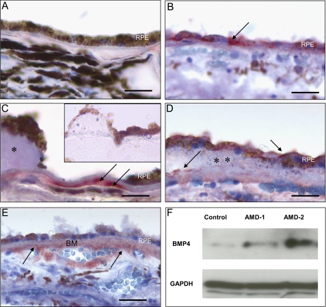
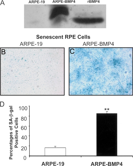
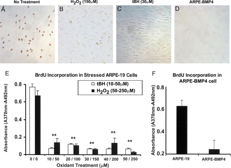
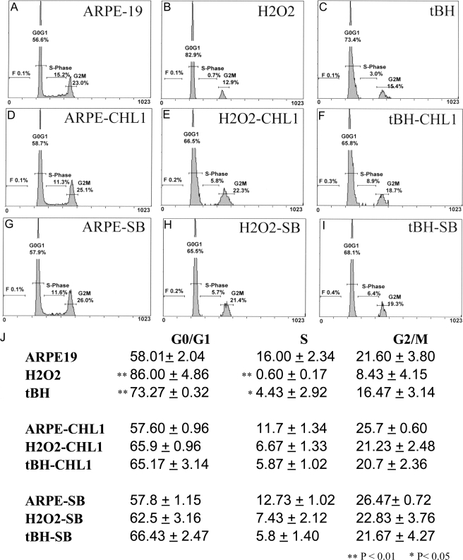
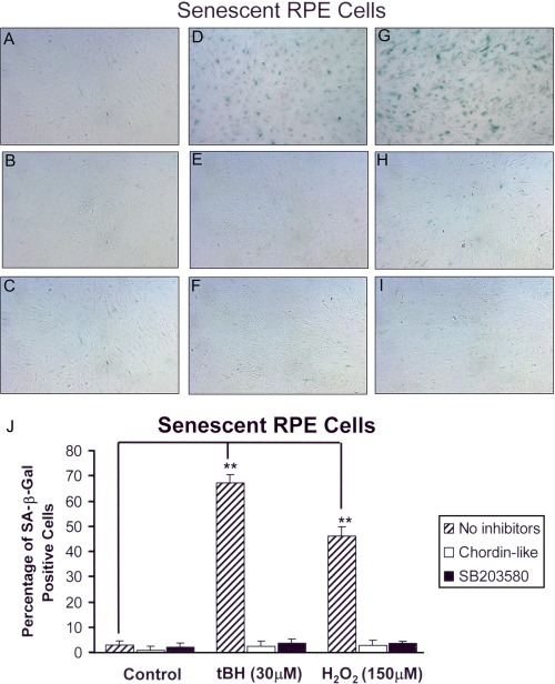
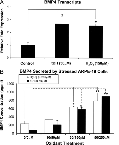
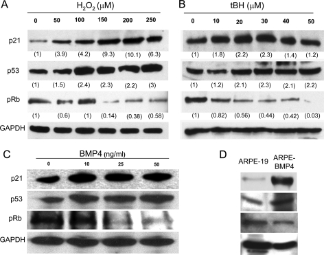
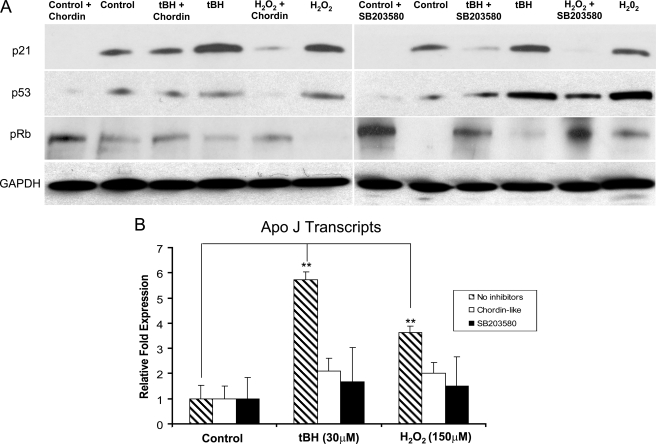
The retinal pigment epithelium is a primary site of pathology in age-related macular degeneration. Oxidative stress and senescence are both thought to be important mediators of macular degeneration pathogenesis. We demonstrate here that bone morphogenetic protein-4 is highly expressed in the retinal pigment epithelium and adjacent extracellular matrix of patients with dry age-related macular degeneration. In vitro studies revealed that sublethal oxidative stress increased bone morphogenetic protein-4 expression in retinal pigment epithelial cells, and both bone morphogenetic protein-4 and persistent mild oxidative stress can induce retinal pigment epithelial cell senescence through p53-p21(Cip1/WAF1)-Rb pathway. We further demonstrate that bone morphogenetic protein-4 acts as a mediator in oxidative stress-induced senescence and that this mediator function is via Smad and the p38 signaling pathway to increase and activate p53 and p21(Cip1/WAF1) and decrease phospho-Rb. Oxidative stress-induced senescence can be blocked by Chordin-like, an antagonist of bone morphogenetic protein-4, or SB203580, a phospho-p38 inhibitor. Our results suggest that oxidative stress and bone morphogenetic protein-4 may interact to promote retinal pigment epithelial cell senescence and that bone morphogenetic protein-4 may represent a novel therapeutic target to inhibit the progressive effects of oxidative stress and senescence in dry age-related macular degeneration.


Figures

FIGURE 1.
FIGURE 1.
Expression of BMP4 in macular tissues of control and AMD patients. Tissue samples, including RPE and choroid, were dissected from the macular region of control patients (n = 12), patients with early AMD (n = 10), and patients with geographic atrophy (n = 3). Frozen sections were stained immunohistochemically using anti-BMP4 antibody, and localization of BMP4 was identified by the red chromogen AEC. In controls (A), BMP4 immunoreactivity was not found. In early AMD samples (B-D), BMP4 was localized to the cytoplasm of some retinal pigment epithelium (RPE)(B, arrow), in thickened Bruch membrane adjacent to hard druse (C, arrows), and in RPE and Bruch membrane (arrows) adjacent to a large soft druse (D, **). Adjacent to areas of RPE loss in geographic atrophy (E), there was prominent immunoreactivity in RPE, in Bruch membrane (BM), and around choroidal vessels (arrows). Parallel sections stained using antibody preadsorbed with rBMP4 showed no immunoreactivity (C, inset). Immunoperoxidase stain with hematoxylin counterstain is shown. Bar, 50 μm (A, B, and D), 25 μm (C), and 100 μm (E). Western blot (F) of dissected macular tissues from control patients and two patients with early AMD (AMD-1 and AMD-2) revealed increased expression of BMP4 protein in AMD maculas; GAPDH revealed equal loading of the samples.
FIGURE 2.
FIGURE 2.
Induction of senescence in BMP4-overexpressing cells. Secretion of BMP4 by a BMP4-overexpressing cell clone (ARPE-BMP4-4) into cell supernatants was measured by Western blot. A, ARPE-BMP4-4 cells secreted large amounts of BMP4 protein into cell culture medium compared with its parental cell line, ARPE-19. Culture medium spiked with rBMP4 represented the positive control. (ARPE-BMP4-4-derived BMP4 contains a 10-amino acid c-myc tag and migrates at a higher molecular weight than rBMP4). B, C, and D, ARPE-19 cells grown in 1% serum-containing medium for 7 days showed low levels of expression of the senescence marker SA-β-galactosidase; however, expression of SA-β-galactosidase was markedly increased in ARPE-BMP4 cells (**, p < 0.001).
FIGURE 3.
FIGURE 3.
BrdU incorporation in ARPE-19 cells after sublethal exposure to oxidants and in BMP4-overexpressing cells. An in situ BrdUrd incorporation immunoperoxidase assay was used in ARPE-19 cells that were treated with H2O2 (150 μm) for two stresses or tBH (30 μm) for five stresses. A-D, light microscopic images of in situ BrdU immunostaining of ARPE-19 cells. A, unstressed ARPE-19 cells; B, H2O2-stressed ARPE-19 cells; C, tBH-stressed ARPE-19 cells; D, BMP4-overexpressing cells. E and F, a BrdU incorporation ELISA was used in ARPE-19 cells that were treated with H2O2 (50-250 μm) for two stresses or tBH (10-50 μm) for five stresses and in BMP4-overexpressing cells. The rate of BrdU incorporation was significantly decreased in stressed RPE cells compared with control cells (E), even at the lowest dose of oxidants, as well as in BMP4 overexpressing cells (F). **, p < 0.001.
FIGURE 4.
FIGURE 4.
Cell cycle analysis of ARPE-19 cells after oxidant treatment and oxidant plus inhibitors. ARPE-19 cells treated with oxidants alone or co-treated with oxidants and BMP4 antagonist Chordin-like 1 (CHL1) or phospho-p38 inhibitor SB203580 (SB) were stained with propidium iodide. The cell cycle distribution was analyzed by flow cytometry. The percentage of cells in each cell cycle phase (G1/G0, S, and G2/M) was determined by DNA content. A representative experiment is shown in A-I, whereas the mean values of three independent experiments are shown in J. The results (J) showed that treatment with oxidants increased the percentage of cells in G0/G1 phase from 58% (unstressed cells) to 86% (H2O2-stressed cells) and 73% (tBH-stressed cells) and decreased the percentage of cells in S phase from 16% (unstressed cells) to 0.6% (H2O2-stressed cells) and 4.4% (tBH-stressed cells), whereas the percentage of cells in G0/G1 induced by oxidative stress and treated with either Chordin-like 1 or SB203580 was decreased to a level similar to those of unstressed cells. *, p < 0.05; **, p < 0.01.
FIGURE 5.
FIGURE 5.
Senescence-associated β-galactosidase staining of ARPE-19 cells treated with oxidants and inhibitors of BMP4 or p38 signaling. The oxidant stressed with or without inhibitor-treated ARPE-19 cells was incubated with SA-β-galactosidase staining buffer for 24 h at 37 °C. Senescent cells were photographed with a phase-contrast microscope and counted as percentage of SA-β-galactosidase-positive cells. A, ARPE-19 cells without stressors or inhibitors; B, ARPE-19 without stressor but with Chordin-like 1; C, ARPE-19 without stressor but with SB203580; D, ARPE-19 cells treated with 150 μm H2O2; E, ARPE-19 cells treated with 150 μm H2O2 and Chordin-like 1; F, ARPE-19 cells treated with 150 μm H2O2 and SB203580; G, ARPE-19 cells treated with 30 μm tBH; H, ARPE-19 cells treated with 30 μm tBH and Chordinlike 1; I, ARPE-19 cells treated with 30 μm tBH and SB203580. The images shown here demonstrate that stressed cells show higher percentage of senescence (SA-β-galactosidase-positive cells; D and G) than controls (A). In the presence of the BMP4 antagonist, Chordin-like 1, or a phospho-p38 inhibitor, SB203580, in the medium, the numbers of senescent RPE cells were reduced to base-line level (E, H, F, and I). J, tBH and H2O2 induced 65 and 48% cell senescence, respectively, whereas the control and stressor plus inhibitor groups showed less than 5% background cell senescence (**, p < 0.001).
FIGURE 6.
FIGURE 6.
Effect of oxidative stress on BMP4 gene expression and protein secretion in ARPE-19 cells. Real time PCR (A) and ELISA (B) were used to quantify the expression of BMP4 transcripts and secretion of BMP4 protein into cell culture supernatants from stressed and unstressed ARPE-19 cells. BMP4 mRNA expression was about 2.5-fold higher in H2O2-stressed cells and about 3-fold higher in tBH-stressed cells than those in nonstressed ARPE-19 cells. BMP4 secretion into the media from oxidant-treated ARPE cells was also increased in a dose-dependent manner. *, p < 0.05; **, p < 0.01.
FIGURE 7.
FIGURE 7.
Western blot analysis of p21Cip1/WAF1, p53, and phospho-Rb proteins in BMP4 or oxidant-treated ARPE-19 cells. A, results using H2O2-treated cells; B, results using tBH-treated cells; C, results using BMP4-treated cells; D, results using BMP4 overexpressing clone ARPE-BMP4. p21Cip1/WAF1 and p53 proteins were increased and phospho-Rb (pRb) protein was decreased in BMP4- and oxidant-treated ARPE-19 cells and in BMP4-overexpressing cells. For oxidant-treated cells, corresponding expression levels were measured by Scion software, normalized to GAPDH, and labeled under each corresponding band.
FIGURE 8.
FIGURE 8.
Western blot analysis of phospho-Smad1/5/8 and phospho-p38 proteins in BMP4- or oxidant-treated ARPE-19 cells. ARPE-19 cells were treated with recombinant BMP4 protein for 1 h, and two oxidants, 150 μm H2O2 for two stresses and 30 μm tBH for five stresses. Western blots of cell lysates were evaluated using phospho-Smad1/5/8 or phospho-p38 antibodies and total Smad1 or total p38 antibodies for normalization. Phospho-Smad1/5/8 and phospho-p38 proteins were increased in BMP4- and oxidant-treated RPE cells.
FIGURE 9.
FIGURE 9.
Effect of Chordin-like 1 and SB203580 on expression of p21Cip1/WAF1, p53, phospho-Rb, and Apo J. A, Western blot analysis of the protein levels of p21Cip1/WAF1, p53, and phospho-Rb (pRb) in oxidant-treated ARPE-19 cells, with or without Chordin-like 1 or SB203580. The higher levels of p21Cip1/WAF1 and p53 protein induced by oxidants were reduced by Chordin-like 1 or SB203580 treatment. In each experiment, equal loading was demonstrated by immunoblot for GAPDH. B, analysis of the expression of senescence marker gene, Apo J, in oxidant treated ARPE-19 cells with or without inhibitors using real time PCR. The expression of Apo J was about 6-fold higher in tBH-treated cells and about 4-fold higher in H2O2-treated cells than the expression in nonstressed control ARPE-19 cells. The increased expression of Apo J in stressed ARPE-19 cells was markedly suppressed when Chordin-like 1 or SB203580 was added into the medium. **, p < 0.01.

References

    1. de Jong, P. T. (2006) N. Engl. J. Med. 355 1474-1485 - PubMed
    1. Gehrs, K. M., Anderson, D. H., Johnson, L. V., and Hageman, G. S. (2006) Ann. Med. 38 450-471 - PMC - PubMed
    1. Klein, R., Klein, B. E., Jensen, S. C., and Meuer, S. M. (1997) Ophthalmology 104 7-21 - PubMed
    1. Ambati, J., Ambati, B. K., Yoo, S. H., Ianchulev, S., and Adamis, A. P. (2003) Surv. Ophthalmol. 48 257-293 - PubMed
    1. Zarbin, M. A. (2004) Arch. Ophthalmol. 122 598-614 - PubMed

Publication types

Substances