. 2009 Mar 20;284(12):7436-45.
doi: 10.1074/jbc.M808999200. Epub 2009 Jan 21.

HDAC6 modulates Hsp90 chaperone activity and regulates activation of aryl hydrocarbon receptor signaling

Affiliations

HDAC6 modulates Hsp90 chaperone activity and regulates activation of aryl hydrocarbon receptor signaling

Vikram D Kekatpure et al. J Biol Chem. .

Retraction in

Abstract

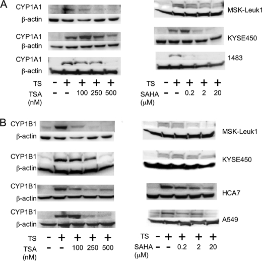
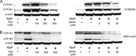
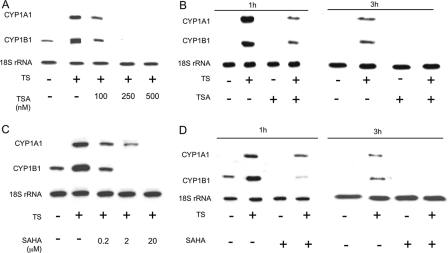
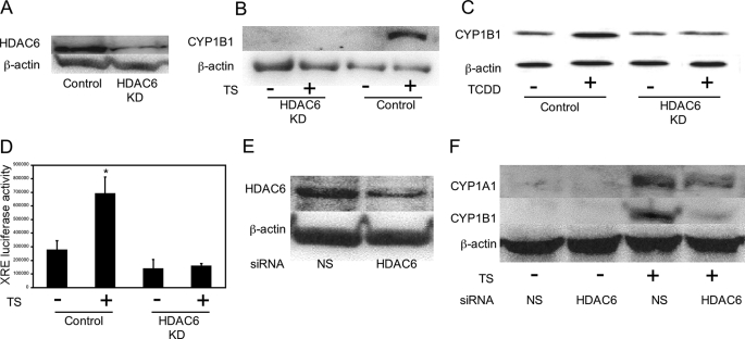
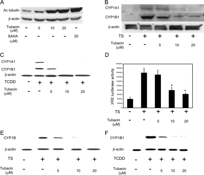
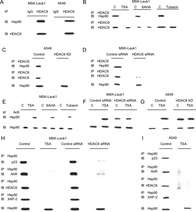
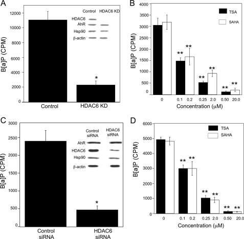
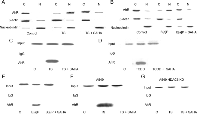
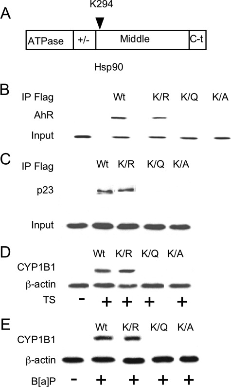
The aryl hydrocarbon receptor (AhR), a ligand-activated member of the basic helix-loop-helix family of transcription factors, binds with high affinity to polycyclic aromatic hydrocarbons (PAH) and the environmental toxin 2,3,7,8-tetrachlorodibenzo-p-dioxin (dioxin). Most of the biochemical, biological, and toxicological responses caused by exposure to PAHs and polychlorinated dioxins are mediated, at least in part, by the AhR. The AhR is a client protein of Hsp90, a molecular chaperone that can be reversibly acetylated with functional consequences. The main objective of this study was to determine whether modulating Hsp90 acetylation would affect ligand-mediated activation of AhR signaling. Trichostatin A and suberoylanilide hydroxamic acid, two broad spectrum HDAC inhibitors, blocked PAH and dioxin-mediated induction of CYP1A1 and CYP1B1 in cell lines derived from the human aerodigestive tract. Silencing HDAC6 or treatment with tubacin, a pharmacological inhibitor of HDAC6, also suppressed the induction of CYP1A1 and CYP1B1. Inhibiting HDAC6 led to hyperacetylation of Hsp90 and loss of complex formation with AhR, cochaperone p23, and XAP-2. Inactivation or silencing of HDAC6 also led to reduced binding of ligand to the AhR and decreased translocation of the AhR from cytosol to nucleus in response to ligand. Ligand-induced recruitment of the AhR to the CYP1A1 and CYP1B1 promoters was inhibited when HDAC6 was inactivated. Mutation analysis of Hsp90 Lys(294) shows that its acetylation status is a strong determinant of interactions with AhR and p23 in addition to ligand-mediated activation of AhR signaling. Collectively, these results show that HDAC6 activity regulates the acetylation of Hsp90, the ability of Hsp90 to chaperone the AhR, and the expression of AhR-dependent genes. Given the established link between activation of AhR signaling and xenobiotic metabolism, inhibitors of HDAC6 may alter drug or carcinogen metabolism.


Figures

FIGURE 1.
FIGURE 1.
HDAC inhibitors suppress TS-mediated induction of CYP1A1 and CYP1B1 in human aerodigestive epithelial cells. In A and B, MSK-Leuk1, KYSE450, 1483, A549, and HCA7 cells were pretreated with indicated concentrations of TSA, SAHA or vehicle for 2 h. Subsequently, the cells were treated with vehicle or TS for 5 h. Cellular lysate protein was then isolated and loaded (100μg/lane) on a 10% SDS-polyacrylamide gel, electrophoresed, and subsequently transferred onto nitrocellulose as described under “Experimental Procedures.” The immunoblot was probed with antibodies specific for CYP1A1 (A), CYP1B1 (B), or β-actin.
FIGURE 2.
FIGURE 2.
HDAC inhibitors suppress B[a]P-mediated induction of CYP1A1 and CYP1B1 in human aerodigestive epithelial cells. KYSE450 and MSK-Leuk1 cells were pretreated with indicated concentrations of TSA, SAHA, or vehicle for 2 h. Subsequently, the cells were exposed to vehicle or 1 μm B[a]P for 5 h. Cellular lysate protein was then isolated and loaded (100 μg) on a 10% SDS-polyacrylamide gel, electrophoresed, and subsequently transferred onto nitrocellulose as described under “Experimental Procedures.” The immunoblot was probed with antibodies specific for CYP1A1 (A), CYP1B1 (B), or β-actin.
FIGURE 3.
FIGURE 3.
HDAC inhibitors suppress TS-mediated induction of CYP1A1 and CYP1B1 mRNA levels. In A and C, MSK-Leuk1 cells were pretreated with indicated concentrations of TSA (A), SAHA (C), or vehicle for 2 h. The cells were then treated with vehicle or TS for an additional 3 h prior to RNA isolation. In B and D, the cells were pretreated with vehicle, TSA (500 nm), or SAHA (20 μm) for 2 h before receiving TS or vehicle for an additional 1 or 3 h prior to RNA isolation. Total RNA was prepared, and 10 μg/lane of RNA was subjected to Northern blotting. The blots were hybridized sequentially with the indicated probes.
FIGURE 4.
FIGURE 4.
HDAC6 is important for activation of AhR signaling. In AD, A549 cells stably expressing pSuper control plasmid (Control) or HDAC6 small hairpin RNA (HDAC6 KD) were used. A, immunoblot analysis of HDAC6 in lysates from A549 Control versus A549 HDAC6 KD cells. B, control or HDAC6 KD cells were treated with vehicle or TS for 5 h. C, control or HDAC6 KD cells were treated with vehicle or 1 nm TCDD for 5 h. D, control or HDAC6 KD cells were transfected with 1.8 μg of a XRE luciferase construct and 0.2 μg of pSVβgal. Thirty-six h after transfection, the cells were treated with vehicle or TS for another 12 h. XRE luciferase activity represents data that have been normalized to β-galactosidase activity. The data are plotted as the means ± S.D. (n = 6). *, p < 0.01 compared with vehicle treated cells. In E and F, MSK-Leuk1 cells were treated with nonspecific (NS) siRNA or HDAC6 siRNA. F, cells were treated with vehicle or TS for 5 h. In A–C, E, and F, 100 μg of cellular lysate protein/lane was loaded onto a 10% SDS-polyacrylamide gel, electrophoresed, and subsequently transferred onto nitrocellulose. The immunoblots were probed with the indicated antibodies.
FIGURE 5.
FIGURE 5.
Tubacin, an HDAC6 inhibitor, suppresses ligand-mediated activation of AhR signaling. A, MSK-Leuk1 cells were treated with indicated concentrations of tubacin or SAHA for 1 h. B and C, MSK-Leuk1 cells were pretreated with indicated concentrations of tubacin or vehicle for 2 h. The cells were then treated with vehicle, TS (B), or 1 nm TCDD (C) for 5 h. D, MSK-Leuk1 cells were transfected with 1.8 μg of a XRE luciferase construct and 0.2 μg pSVβ-gal. Thirty-six h after transfection, the cells were treated with vehicle or the indicated concentration of tubacin for 2 h. The cells were then treated with vehicle or TS for an additional 12 h. Luciferase activity represents data that have been normalized to β-galactosidase activity. The data are plotted as the means ± S.D. (n = 6). *, p < 0.01 compared with TS-treated cells. E and F, A549 cells were pretreated with vehicle or the indicated concentration of tubacin for 2 h. The cells were then treated with vehicle, TS (E), or TCDD (F) for 5 h. In A–C, E, and F, cell lysates were prepared and loaded (100 μg/lane) on a 10% SDS-polyacrylamide gel, electrophoresed, and subsequently transferred onto nitrocellulose. Immunoblots were probed with the indicated antibodies.
FIGURE 6.
FIGURE 6.
HDAC6 associates with Hsp90 and regulates its acetylation and chaperone complex formation with p23 and XAP-2. A, cell lysates (250 μg) from MSK-Leuk1 and A549 cells were subjected to immunoprecipitation (IP) with antibody to HDAC6 or IgG. The immunoprecipitates were then subjected to immunoblotting (IB) and probed with antibodies to Hsp90 or HDAC6. B, MSK-Leuk1 cells were treated with vehicle (lanes C), TSA (500 nm), SAHA (20 μm), or tubacin (20 μm) for 1 h. The cell lysates were prepared and immunoprecipitated with an antibody to HDAC6. Immunoprecipitates were then subjected to immunoblotting and probed with antibodies to Hsp90 or HDAC6. C, cell lysates were prepared from A549 cells stably expressing pSUPER (Control) or HDAC6 siRNA (HDAC6 KD). D, cell lysates were prepared from MSK-Leuk1 cells transiently transfected with nonspecific (control) siRNA or HDAC6 siRNA. In C and D, cell lysates were subjected to immunoprecipitation with antibody to HDAC6. The immunoprecipitates were then subjected to immunoblotting and probed for Hsp90 and HDAC6. Additionally, cell lysates were directly subjected to immunoblotting for Hsp90. In EG, MSK-Leuk1 and A549 cells were treated as indicated with vehicle (lanes C), TSA (500 nm), SAHA (20 μm), or tubacin (20 μm) for 1 h. The cell lysates were then subjected to immunoprecipitation with antibody to acetyl lysine (AcK). The immunoprecipitates were then subjected to immunoblotting and probed for Hsp90. Cell lysates were also directly subjected to immunoblotting for Hsp90. H, cell lysates were prepared from MSK-Leuk1 cells that received vehicle (control) or TSA (500 nm) for 1 h or were transfected with nonspecific siRNA (control siRNA) or HDAC6 siRNA. I, cell lysates were prepared from A549 cells that received vehicle or TSA (500 nm) for 1 h. In H and I, cell lysates were subjected to immunoprecipitation with antibody to Hsp90. The immunoprecipitates were then subjected to immunoblotting and probed for p23, AhR, HDAC6, and XAP-2. Additionally, cell lysates were directly subjected to immunoblotting for Hsp90.
FIGURE 7.
FIGURE 7.
HDAC6 is necessary for optimal binding of ligand to AhR. In AD, cytosols were isolated from cells and incubated overnight at 4 °C with 100 nm [3H]B[a]P. The ligand binding assay was performed as described under “Experimental Procedures.” Binding of B[a]P is expressed as cpm of [3H]B[a]P/100 μl of cell cytosol. A, cytosols from A549 cells stably expressing pSUPER control plasmid (Control) or HDAC6 siRNA (HDAC6 KD) were subjected to ligand binding assay. Inset, cell lysates were subjected to immunoblotting and probed for HDAC6, AhR, Hsp90, and β-actin. B, cytosols were prepared from A549 cells that were treated with the indicated concentrations of TSA and SAHA for 1 h prior to ligand binding assay. C, cytosols were prepared from MSK-Leuk1 cells that were transiently transfected with control siRNA or HDAC6 siRNA prior to ligand binding assay. Inset, cell lysates were subjected to immunoblotting and probed as indicated. D, cytosols were prepared from MSK-Leuk1 cells that were treated with the indicated concentrations of TSA and SAHA for 1 h prior to ligand binding assay. The data are plotted as the means ± S.D. (n = 3). *, p < 0.01; **, p < 0.001. The data shown are representative of three independent experiments.
FIGURE 8.
FIGURE 8.
HDAC inhibitors block PAH induced translocation of AhR from the cytosol to the nucleus and activation of CYP1A1 and CYP1B1 transcription. MSK-Leuk1 (AE) cells and A549 (F and G) cells were pretreated with vehicle or 20 μm SAHA for 2 h. Subsequently, the cells were treated as indicated with vehicle, TS, B[a]P (1 μm), or TCDD (10 nm). In A and B, the cells were lysed, nuclear (lanes N) and cytosolic (lanes C) fractions were isolated, and protein (100 μg/lane) was subjected to immunoblotting. The immunoblots were probed with the indicated antibodies. In CG, ChIP assays were performed. Chromatin fragments were immunoprecipitated with antibodies against AhR, and the CYP1A1 (CE) and CYP1B1 promoters (F and G) were amplified by PCR. DNA sequencing was carried out, and the PCR product was confirmed to be the correct promoter. The CYP1A1 and CYP1B1 promoters were not detected when normal IgG was used or when antibody was omitted from the immunoprecipitation step (data not shown).
FIGURE 9.
FIGURE 9.
Lys294 is important for Hsp90 interaction with AhR. A represents the structure of Hsp90 protein. B and C, A549 cells were stably transfected with the indicated FLAG-Hsp90 constructs. The cells were lysed, and FLAG-Hsp90 immunoprecipitates (IP) were then subjected to immunoblotting and probed for AhR (B) and p23 (C). In D and E, the cells transfected with the indicated FLAG-Hsp90 constructs were treated for 5 h with vehicle, TS, or B[a]P (1μm). Following treatment, cell lysates (100μg/lane) were subjected to immunoblotting for CYP1B1 and β-actin, respectively.

Comment in

  • Findings of Research Misconduct.
    [No authors listed] [No authors listed] Fed Regist. 2023 Sep 13;88(176):62800-62803. Fed Regist. 2023. PMID: 37736072 Free PMC article. No abstract available.
  • Findings of Research Misconduct.
    [No authors listed] [No authors listed] Fed Regist. 2023 Sep 13;88(176):62803-62807. Fed Regist. 2023. PMID: 37736073 Free PMC article. No abstract available.

References

    1. Gu, Y. Z., Hogenesch, J. B., and Bradfield, C. A. (2000) Annu. Rev. Pharmacol. Toxicol. 40 519–561 - PubMed
    1. Poland, A., Glover, E., and Kende, A. S. (1976) J. Biol. Chem. 251 4936–4946 - PubMed
    1. Bock, K. W., and Köhle, C. (2006) Biochem. Pharmacol. 72 393–404 - PubMed
    1. Andersson, P., McGuire, J., Rubio, C., Gradin, K., Whitelaw, M. L., Pettersson, S., Hanberg, A., and Poellinger, L. (2002) Proc. Natl. Acad. Sci. U. S. A. 99 9990–9995 - PMC - PubMed
    1. Moennikes, O., Loeppen, S., Buchmann, A., Andersson, P., Ittrich, C., Poellinger, L., and Schwarz, M. (2004) Cancer Res. 64 4707–4710 - PubMed

Publication types

MeSH terms

Substances

LinkOut - more resources