Comparative Study
. 2009 Apr;83(7):3249-57.
doi: 10.1128/JVI.01721-08. Epub 2009 Jan 21.

Robust in vivo transduction of a genetically stable Epstein-Barr virus episome to hepatocytes in mice by a hybrid viral vector

Affiliations
Comparative Study

Robust in vivo transduction of a genetically stable Epstein-Barr virus episome to hepatocytes in mice by a hybrid viral vector

Sean D Gallaher et al. J Virol. 2009 Apr.

Abstract

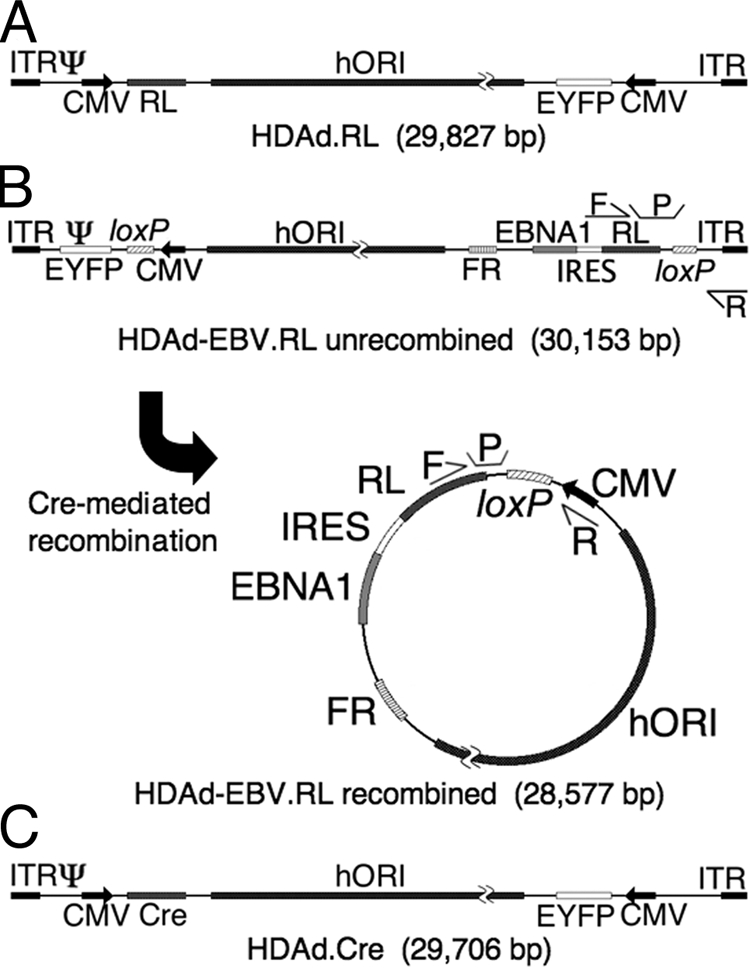
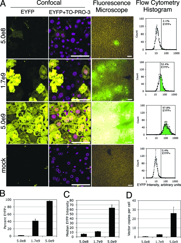
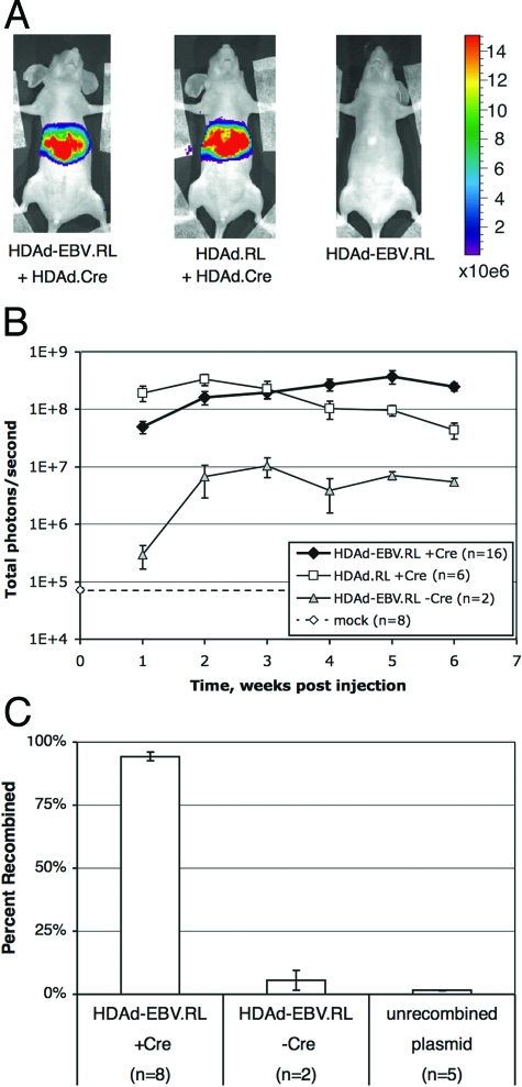
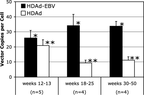
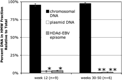
To make a safe, long-lasting gene delivery vehicle, we developed a hybrid vector that leverages the relative strengths of adenovirus and Epstein-Barr virus (EBV). A fully gene-deleted helper-dependent adenovirus (HDAd) is used as the delivery vehicle for its scalability and high transduction efficiency. Upon delivery, a portion of the HDAd vector is recombined to form a circular plasmid. This episome includes two elements from EBV: an EBV nuclear antigen 1 (EBNA1) expression cassette and an EBNA1 binding region. Along with a human replication origin, these elements provide considerable genetic stability to the episome in replicating cells while avoiding insertional mutagenesis. Here, we demonstrate that this hybrid approach is highly efficient at delivering EBV episomes to target cells in vivo. We achieved nearly 100% transduction of hepatocytes after a single intravenous injection in mice. This is a substantial improvement over the transduction efficiency of previously available physical and viral methods. Bioluminescent imaging of vector-transduced mice demonstrated that luciferase transgene expression from the hybrid was robust and compared well to a traditional HDAd vector. Quantitative PCR analysis confirmed that the EBV episome was stable at approximately 30 copies per cell for up to 50 weeks and that it remained circular and extrachromosomal. Approaches for adapting the HDAd-EBV hybrid to a variety of disease targets and the potential benefits of this approach are discussed.


Figures

FIG. 1.
FIG. 1.
Structure of HDAd vectors. Three vectors were used in the course of these experiments. Each contains an EYFP gene expressed from a constitutive human CMV major immediate-early promoter (CMV); a 19-kb region of human genomic DNA from chromosome 10 for use as a stuffer and as an origin of replication (hORI) (21); and two regions from adenovirus, the inverted terminal repeats (ITR) that function as replication origins and the viral packaging signal (Ψ) that are required in cis for vector packaging and amplification. (A) HDAd.RL is a traditional HDAd vector that was used as a control in these experiments. It expresses RL constitutively from a CMV promoter. (B) HDAd-EBV.RL is a hybrid vector that is designed to deliver a circular EBV episome into target cells by means of a linear HDAd genome. In its linear form, the EBV episome elements are flanked by parallel recognition sites for Cre recombinase (loxP). In the presence of Cre recombinase, the intervening region is excised and circularized as shown. This places a CMV promoter upstream of an expression cassette that contains the RL transgene, an encephalomyocarditis virus internal ribosomal entry site (IRES), and the EBNA1 gene. The episome also caries the FR region that functions with EBNA1 protein to provide a maintenance function to the episome in replicating cells. The relative location of two sets of qPCR primers (F and R) and probes (P) are indicated. One set flanks one of the loxP sites in the linear conformation of the hybrid vector, and a second set flanks the loxP site in the circularized conformation. These were used in qPCR experiments to determine the relative fraction of recombined and unrecombined vector. (C) HDAd.Cre is a traditional HDAd vector that is designed to express Cre recombinase in target cells by means of a CMV promoter. Coinfection of a target cell by both HDAd-EBV.RL and HDAd.Cre is required to form the circular EBV episome.
FIG. 2.
FIG. 2.
Efficiency of hepatocyte transduction in vivo. Adult, female nude mice were injected with HDAd-EBV.RL via the tail vein. Two mice at each of three different vector doses (5.0 × 10⁸, 1.7 × 10⁹, and 5.0 × 10⁹ IGU per mouse as indicated) and a mock-infected mouse were compared. At 6 days postinjection, the mice were sacrificed and liver tissue was harvested for the following analyses. Expression of the EYFP transgene from the vector was used to determine the efficiency of liver transduction. (A) Representative samples of liver tissue from each vector dose are presented. The first and second columns are laser scanning confocal micrographs of 50-μm-thick sections of fixed liver tissue. EYFP indicates vector-transduced cells. TO-PRO-3 was used as a nuclear stain. The third column contains micrographs taken from an epifluorescence- light microscope of 5-μm-thick unfixed sections. Signal intensity was adjusted for image quality in the confocal micrographs but is consistent across all of the fluorescent micrographs. Size bar equals 50 μm in all micrographs. The fourth column displays histograms of EYFP intensity quantified by analytical flow cytometry of single cell suspensions of liver tissue. The vertical line indicates the positive/negative threshold of EYFP as determined by the mock control samples. (B) Aggregate data from the flow cytometry analysis is presented. Each bar represents the mean percentage of EYFP-positive cells at each dose from replicate samplings (n = 3). (C) Additional aggregate cytometry data from the samples described above demonstrate the median signal intensity of EYFP-positive cells as determined from the cytometry histograms. Each bar represents the mean of replicate samplings (n = 3). (D) The number of vector genomes per diploid cell was quantified by qPCR. Each bar represents the mean of replicate samplings (n = 5). Error bars indicate mean ± the SEM.
FIG. 3.
FIG. 3.
EBV episome delivery by HDAd-EBV vectors. Adult, female nude mice were injected intravenously with HDAd vectors and assayed for RL expression by bioluminescent imaging. Four cohorts were compared. Group 1 received 5 × 10⁹ IGU of HDAd-EBV.RL and 5 × 10⁹ IGU of HDAd.Cre (n = 16). Group 2 received 5 × 10⁹ IGU of HDAd.RL and 5 × 10⁹ IGU of HDAd.Cre (n = 6). Group 3 received 1010 IGU of HDAd-EBV.RL (n = 2). To determine the background of the assay, a fourth “mock” group was imaged using the same conditions as the experimental groups but received no vector (n = 8). (A) Representative images from the first three groups at 1 week postinfection. The total number of photons per second, as averaged over a 5-min bioluminescent exposure, was overlaid on an optical image of the mouse. The signal intensity was color coded according to the included scale from 1.5 × 10⁵ to 1.5 × 10⁷ total photons/s. (B) Mice were imaged weekly over a 6-week period after vector injection as in panel A. The number of photons/second detected in the region of the liver was plotted for each cohort over time. The dashed line indicates the background of the assay as determined by the mock cohort. Error bars indicate the mean ± the SEM. (C) Total DNA was prepared from samples of transduced liver tissue for analysis of recombination at the loxP sites by qPCR. Samples ranged from 9 to 30 weeks postinjection. A total of 10 mice injected with HDAd-EBV.RL were analyzed. Eight received HDAd.Cre, and two received no Cre. Each result is presented as a percentage of recombined vector relative to the total. A plasmid with only the unrecombined sequence was used to determine the background of the assay. Error bars indicate the mean ± the SEM.
FIG. 4.
FIG. 4.
Persistence of viral DNA in vivo. Liver tissue was harvested at various time points from adult, female nude mice cotransduced with 5 × 10⁹ IGU of HDAd-EBV.RL and 5 × 10⁹ IGU of HDAd.Cre. Vector DNA in each tissue sample was quantified by qPCR with a primer-probe set unique to each vector. Values were normalized to the number of murine cells per sample as quantified by a third primer-probe set specific to the mouse genome. To facilitate the evaluation of the data, samples were divided into three groups based on the number of weeks postinjection that the sample was harvested. n, Number of mice in each group. Error bars represent the mean ± the SEM. Variation between the three time frames was determined by one-way Fisher analysis of variance. There was no statistically significant change in the HDAd-EBV hybrid vector (*, P = 0.52), but there was a significant decrease in the linear HDAd vector (**, P = 0.04).
FIG. 5.
FIG. 5.
Integration state of vector DNA. A selective salt precipitation was performed to separate the DNA from vector-transduced liver tissue into HMW and LMW fractions. A 35-kb plasmid was added to each sample prior to precipitation as a control for separation of HMW and LMW DNA. The number of copies of chromosomal DNA, plasmid DNA, and HDAd-EBV vector DNA in each fraction were assayed in separate qPCR reactions. Each bar represents the percentage of copies in the high-molecular-weight fraction relative to the total copy number from both fractions. “Week 12” represents nine preparations of DNA from three mice harvested 12 weeks postinjection. “Weeks 30 to 50” represents six preparations from three mice harvested during the later time period. Error bars indicate mean ± the SEM. Variation between the plasmid DNA and HDAd-EBV episome DNA was insignificant as determined by one-way Fisher analysis of variance (*, P = 0.50; **, P = 0.13).

References

    1. Berry, M. N., and A. M. Edwards (ed.). 2000. The hepatocyte review. Kluwer Academic Publishers, Boston, MA.
    1. Bhaumik, S., and S. S. Gambhir. 2002. Optical imaging of Renilla luciferase reporter gene expression in living mice. Proc. Natl. Acad. Sci. USA 99377-382. - PMC - PubMed
    1. Bloquel, C., E. Fabre, M. F. Bureau, and D. Scherman. 2004. Plasmid DNA electrotransfer for intracellular and secreted proteins expression: new methodological developments and applications. J. Gene Med. 6(Suppl. 1)S11-S23. - PubMed
    1. Brauner, R., M. Nonoyama, H. Laks, D. C. Drinkwater, Jr., S. McCaffery, T. Drake, A. J. Berk, L. Sen, and L. Wu. 1997. Intracoronary adenovirus-mediated transfer of immunosuppressive cytokine genes prolongs allograft survival. J. Thorac. Cardiovasc. Surg. 114923-933. - PubMed
    1. Brooks, A. R., R. N. Harkins, P. Wang, H. S. Qian, P. Liu, and G. M. Rubanyi. 2004. Transcriptional silencing is associated with extensive methylation of the CMV promoter following adenoviral gene delivery to muscle. J. Gene Med. 6395-404. - PubMed

Publication types