. 2009 Apr;83(7):3200-11.
doi: 10.1128/JVI.02205-08. Epub 2009 Jan 21.

Proteolytic activation of the 1918 influenza virus hemagglutinin

Affiliations

Proteolytic activation of the 1918 influenza virus hemagglutinin

Chawaree Chaipan et al. J Virol. 2009 Apr.

Abstract

Proteolytic activation of the hemagglutinin (HA) protein is indispensable for influenza virus infectivity, and the tissue expression of the responsible cellular proteases impacts viral tropism and pathogenicity. The HA protein critically contributes to the exceptionally high pathogenicity of the 1918 influenza virus, but the mechanisms underlying cleavage activation of the 1918 HA have not been characterized. The neuraminidase (NA) protein of the 1918 influenza virus allows trypsin-independent growth in canine kidney cells (MDCK). However, it is at present unknown if the 1918 NA, like the NA of the closely related strain A/WSN/33, facilitates HA cleavage activation by recruiting the proprotease plasminogen. Moreover, it is not known which pulmonary proteases activate the 1918 HA. We provide evidence that NA-dependent, trypsin-independent cleavage activation of the 1918 HA is cell line dependent and most likely plasminogen independent since the 1918 NA failed to recruit plasminogen and neither exogenous plasminogen nor the presence of the A/WSN/33 NA promoted efficient cleavage of the 1918 HA. The transmembrane serine protease TMPRSS4 was found to be expressed in lung tissue and was shown to cleave the 1918 HA. Accordingly, coexpression of the 1918 HA with TMPRSS4 or the previously identified HA-processing protease TMPRSS2 allowed trypsin-independent infection by pseudotypes bearing the 1918 HA, indicating that these proteases might support 1918 influenza virus spread in the lung. In summary, we show that the previously reported 1918 NA-dependent spread of the 1918 influenza virus is a cell line-dependent phenomenon and is not due to plasminogen recruitment by the 1918 NA. Moreover, we provide evidence that TMPRSS2 and TMPRSS4 activate the 1918 HA by cleavage and therefore may promote viral spread in lung tissue.


Figures
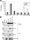
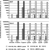
FIG. 1.
FIG. 1.
Trypsin activation of 1918 influenza virus HA pseudotypes is required for infectivity independent of the presence of NA. (A) 293T cells were infected with env-defective, p24-normalized HIV-1 NL4-3 reporter virus pseudotyped with the indicated glycoproteins. Prior to infection, the virions were treated with trypsin or PBS, and after 10 min soybean trypsin inhibitor was added. Three days after infection, luciferase activities in cellular lysates were determined. Infection observed with glycoprotein harboring pseudotypes is shown relative to infection detected with control particles bearing no glycoprotein. The average of four independent experiments with different virus stocks is shown. Error bars indicate standard errors of the means. (B) HIV-1-based VLPs with the indicated combinations of surface proteins were generated in 293T cells and concentrated by centrifugation through a 20% sucrose gradient. The VLP preparations were normalized for comparable content of HIV-1 Gag (p55), pretreated with trypsin or PBS, separated by SDS-polyacrylamide gel electrophoresis, and analyzed by Western blotting. The 1918 influenza virus HA was detected with a mouse monoclonal antibody. Similar results were obtained in two independent experiments with different VLP preparations.
FIG. 2.
FIG. 2.
Impact of spinoculation and protease inhibitors on 1918 influenza virus HA-driven viral entry. (A) 293T, Huh-7, and MDCK cells were seeded in 96-well plates and infected with p24-normalized pseudotypes bearing 1918 influenza virus HA, NA, and M2 or VSV-G. The virions were pretreated with trypsin or PBS and, subsequently, a trypsin inhibitor, before addition to target cells. Two plates were infected in parallel: one plate was incubated at 37°C immediately after addition of virus; the other plate was centrifuged for 2 h at 25°C and 1,200 rpm after infection and then incubated at 37°C. Three days after infection, luciferase activities in cellular lysates were determined. A representative experiment performed in triplicates is shown. Error bars indicate standard deviations. Similar results were obtained in two independent experiments. (B) Huh-7 cells were incubated with the indicated concentrations of the cysteine protease inhibitor E64d prior to infection with infectivity-normalized pseudotypes carrying the 1918 influenza virus HA, NA, M2, ZEBOV-GP, or VSV-G. Pseudotypes bearing influenza virus proteins were pretreated with PBS or trypsin for 10 min at room temperature, and soybean trypsin inhibitor was added. Three days after infection, luciferase activities in cellular lysates were determined. Infection of Huh-7 cells in the absence of protease inhibitors was set as 100%. The average of three independent experiments performed in triplicate is shown. Error bars indicate standard errors of the means. cps, counts per second.
FIG. 3.
FIG. 3.
A/WSN/33 NA fails to promote cleavage activation of the 1918 HA despite efficient recruitment of plasminogen. (A) Pseudotypes bearing the indicated glycoproteins were PBS or trypsin treated and used for infection of Huh-7 cells. Three days after infection, luciferase activities in cellular lysates were determined. The results of a representative experiment are shown and were confirmed in two independent experiments. Error bars indicate standard deviations. (B) VLPs bearing the 1918 HA and NA were incubated with PBS, trypsin, or plasminogen, and the 1918 HA was visualized by Western blotting. (C) The experiment was carried out as described for panel A. However, pseudotypes were treated with PBS, trypsin, or plasminogen (100 μg/ml). The results of a representative experiment are presented and were confirmed within a separate experiment. (D) The indicated proteins were transiently expressed in 293T cells, the transfected cells were incubated with PBS or purified plasminogen (10 μg/ml), and plasminogen binding was detected by FACS. A representative experiment is shown. Similar results were obtained in two independent experiments. PMG, plasminogen; cps, counts per second.
FIG. 4.
FIG. 4.
Lysates from MDCK but not 293T cells activate 1918 HA-driven viral entry. Pseudotypes bearing the 1918 influenza virus HA, NA, and M2, or VSV-G were pretreated with trypsin and, subsequently, soybean trypsin inhibitor or lysates from MDCK or 293T cells. After 30 min the treated pseudotypes were used for infection of 293T and Huh-7 cells. Three days after infection, luciferase activities in cellular lysates were determined. The results of a representative experiment performed in triplicate are shown and were confirmed in two separate experiments. Error bars indicate standard deviations. Statistical significance was calculated employing a two-tailed t test for independent samples. cps, counts per second.
FIG. 5.
FIG. 5.
The 1918 influenza virus replicates efficiently in MDCK but not Huh-7 cells in the absence of trypsin. MDCK and Huh-7 cells were infected at an MOI of 0.001 and then cultivated in the absence or presence of trypsin. Supernatants were collected at 24, 48, and 72 h p.i. from MDCK cultures and at 48 h p.i. from Huh-7 cultures, and the viral titers were determined by a 50% tissue culture infective dose (TCID50) assay on fresh MDCK cells. A representative experiment performed in duplicate is shown; error bars indicate standard deviations.
FIG. 6.
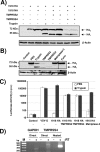
FIG. 6.
TMPRSS2 and TMPRSS4 activate 1918 HA by cleavage. (A) 293T cells were transiently cotransfected with the 1918 HA (jointly with pcDNA3 or the 1918 NA) and TMPRSS2, TMPRSS4, or mouse matriptase-3 (HA and protease expression plasmids were transfected at a 3:1 ratio). Subsequently, the transfected cells were treated with trypsin or PBS, and proteolytic processing of HA was analyzed by Western blotting. (B) 293T cells were transfected with the 1918 HA alone or in combination with the indicated proteases (HA and protease expression plasmids were transfected at a 1:1 ratio); the cells were treated with PBS or trypsin, and proteolytic processing of HA was analyzed by Western blotting. (C) Pseudotypes were generated in 293T cells expressing the empty vector or the indicated proteases (HA and protease expression plasmids were transfected at a 3:1 ratio), normalized to p24 (150 pg/well), and employed for infection of Huh-7 cells. Three days after infection, luciferase activities in cellular lysates were determined. A representative experiment is shown, and similar results were obtained in two independent experiments. Error bars indicate standard deviations. (D) RNA was obtained from cells present in bronchoalveolar lavage fluids and reverse transcribed within reaction mixtures containing reverse transcriptase (RT) enzyme or PBS, and GAPDH and TMPRSS4 were amplified by PCR. M, molecular weight marker. cps, counts per second.

References

    1. Ahmed, R., M. B. Oldstone, and P. Palese. 2007. Protective immunity and susceptibility to infectious diseases: lessons from the 1918 influenza pandemic. Nat. Immunol. 81188-1193. - PMC - PubMed
    1. Belshe, R. B. 2005. The origins of pandemic influenza—lessons from the 1918 virus. N. Engl. J. Med. 3532209-2211. - PubMed
    1. Betakova, T. 2007. M2 protein—a proton channel of influenza A virus. Curr. Pharm. Des. 133231-3235. - PubMed
    1. Bosch, V., B. Kramer, T. Pfeiffer, L. Starck, and D. A. Steinhauer. 2001. Inhibition of release of lentivirus particles with incorporated human influenza virus haemagglutinin by binding to sialic acid-containing cellular receptors. J. Gen. Virol. 822485-2494. - PubMed
    1. Böttcher, E., T. Matrosovich, M. Beyerle, H. D. Klenk, W. Garten, and M. Matrosovich. 2006. Proteolytic activation of influenza viruses by serine proteases TMPRSS2 and HAT from human airway epithelium. J. Virol. 809896-9898. - PMC - PubMed

Publication types

MeSH terms

LinkOut - more resources