. 2009 Apr;83(8):3877-90.
doi: 10.1128/JVI.01733-08. Epub 2009 Jan 21.

Self-assembly of Epstein-Barr virus capsids

Affiliations

Self-assembly of Epstein-Barr virus capsids

Brandon W Henson et al. J Virol. 2009 Apr.

Abstract

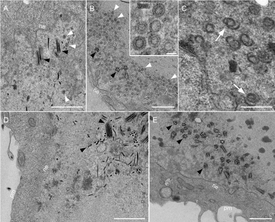
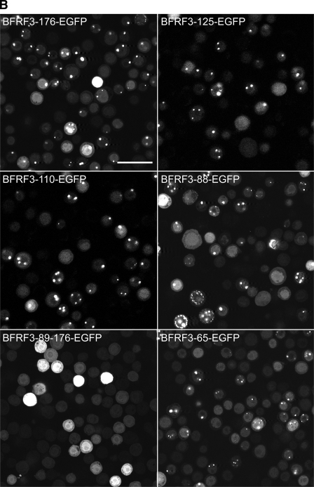
Epstein-Barr virus (EBV), a member of the Gammaherpesvirus family, primarily infects B lymphocytes and is responsible for a number of lymphoproliferative diseases. The molecular genetics of the assembly pathway and high-resolution structural analysis of the capsid have not been determined for this lymphocryptovirus. As a first step in studying EBV capsid assembly, the baculovirus expression vector (BEV) system was used to express the capsid shell proteins BcLF1 (major capsid protein), BORF1 (triplex protein), BDLF1 (triplex protein), and BFRF3 (small capsid protein); the internal scaffold protein, BdRF1; and the maturational protease (BVRF2). Coinfection of insect cells with the six viruses expressing these proteins resulted in the production of closed capsid structures as judged by electron microscopy and sedimentation methods. Therefore, as shown for other herpesviruses, only six proteins are required for EBV capsid assembly. Furthermore, the small capsid protein of EBV (BFRF3), like that of Kaposi's sarcoma-associated herpesvirus, was found to be required for assembly of a stable structure. Localization of the small capsid protein to nuclear assembly sites required both the major capsid (BcLF1) and scaffold proteins (BdRF1) but not the triplex proteins. Mutational analysis of BFRF3 showed that the N-terminal half (amino acids 1 to 88) of this polypeptide is required and sufficient for capsid assembly. A region spanning amino acids 65 to 88 is required for the concentration of BFRF3 at a subnuclear site and the N-terminal 65 amino acids contain the sequences required for interaction with major capsid protein. These studies have identified the multifunctional role of the gammaherpesvirus small capsid proteins.


Figures

FIG. 1.
FIG. 1.
Expression of the EBV capsid proteins in insect cells using recombinant baculoviruses. Sf21 cells (1 × 10⁶) were infected with each baculovirus (50 μl) expressing the six EBV capsid proteins. Infected cells were harvested 72 h after infection, and the extracts prepared were analyzed by SDS-PAGE (15% acrylamide). The gel was stained with Coomassie brilliant blue to reveal the polypeptides. The position of the EBV capsid protein is indicated with an asterisk to the right of each lane. BAC is a baculovirus that does not contain a gene insert, and mock-infected (MI) cells are also shown. The protein standards are in lane M, and the size of each band in kDa is shown to the left.
FIG. 2.
FIG. 2.
Self-assembly of EBV capsids. (A) Sf9 cells were coinfected with all six baculoviruses expressing the EBV capsid proteins, and the infected cells were examined by conventional EM 68 h after infection. Numerous EBV closed capsid structures were evident in the nucleus of the infected cells (white arrowheads). (B) The EBV capsids contained characteristic internal scaffold protein core structures. (C) Sf21 cells were coinfected with baculoviruses expressing all six capsid proteins (EBV-ALL), and the lysates sedimented through sucrose gradients. Reflected light was used to visualize the EBV capsid band. HSV-1 strain KOS-infected cell lysates were also analyzed, and A and B capsids were visible. (D) Material harvested from sucrose gradients was negatively stained and examined by EM. (E) Polypeptide composition of sucrose gradient-purified capsids was examined using a 4 to 12% Nu-Page Bis-Tris gel using MES (morpholineethanesulfonic acid) buffer (Invitrogen). The five abundant capsid proteins are indicated to the right. Protein standards are in lane M, and the sizes in kDa (estimated in MES buffer) of the bands are to the left of the lane. There is usually a small size discrepancy with our protein samples in Nu-Page gels due to our Laemmli buffer. Scale bar, 500 nm; inset bar, 100 nm. The nuclear envelope (ne) and mitochondria (m) are indicated. The locations of baculovirus particles are indicated by black arrowheads in panel A.
FIG. 3.
FIG. 3.
The major capsid and triplex proteins are essential for EBV self-assembly. (A) Sf21 cells were coinfected with all six baculoviruses expressing the EBV capsid proteins, and thin sections were examined by TEM. In similar infections, the virus expressing BVRF2 protease (B to E) was omitted from the infection as well as the virus expressing the BdRF1 scaffold (C), both BORF1 and BDLF1 triplex proteins (D), and the BcLF1-major capsid protein (E). White arrowheads indicate the location of closed EBV capsid structures, and white arrows indicate open capsid shells. Black arrowheads mark baculovirus particles, and the nuclear envelope (ne) and plasma membrane (pm) are indicated where visible. Scale bars, 500 nm; inset bar, 100 nm.
FIG. 4.
FIG. 4.
The EBV small capsid protein (BFRF3) is required for capsid assembly. (A) Sf21 cells were infected with the six baculoviruses expressing EBV capsid proteins (EBV-ALL), and similar infections were carried out in the absence of the virus expressing BFRF3 (−BFRF3). The lysates were sedimented in sucrose gradients and viewed using reflected light. A light-scattering band corresponding to EBV capsids (indicated by arrow) was visible in the EBV-ALL gradient but not in the −BFRF3 gradient. (B) Sf21 cells were coinfected with the baculoviruses expressing BcLF1, BORF1, BDLF1, BdRF1, and BFRF3. Thin sections of infected cells were examined by TEM, and many closed EBV capsid structures were evident in the nucleus (white arrowheads). (C) Similar infections were carried out as described for panel B, except that the virus expressing BFRF3 was left out of the infection. Scale bar, 1,000 nm. The black arrowheads indicate baculovirus particles, and the nuclear envelope (ne) and mitochondria (m) are also marked.
FIG. 5.
FIG. 5.
Localization of BFRF3 to the capsid assembly sites requires both BcLF1 and BdRF1. Sf21 cells were infected with the virus expressing BFRF3-EGFP. In EBV-ALL infections, the viruses expressing BcLF1, BdRF1, BORF1, and BDLF1 were also added to the cells (EBV-ALL). In similar infections, the virus expressing the major capsid protein (−BcLF1), scaffold protein (−BdRF1), triplex 1 protein (−BORF1), or triplex 2 protein (−BDLF1) was left out of the infection. The cells were imaged using a confocal microscope (Zeiss LSM510) 24 h postinfection. The objective was ×40. Scale bar, 50 μm; inset scale bar, 10 μm.
FIG. 6.
FIG. 6.
The assembly domain of BFRF3 maps to the N-terminal half of the polypeptide. (A) The 176-amino-acid sequence of BFRF3 (strain B958) is shown as well as the locations (arrow) of the N-terminal truncation mutants. The C-terminal half of the polypeptide starts at amino acid 89 (gray arrow). (B) Sf21 cells were infected with the baculovriuses expressing HA- or EGFP-tagged BFRF3 mutant polypeptides. Cells were harvested 72 h after infection, and the lysates were analyzed by SDS-PAGE (17% acrylamide for HA or 12% acrylamide for EGFP) and Western blotting methods using anti-HA (αHA) or anti-GFP (αGFP) antibodies. 14C-labeled protein standards are in lane M, and the sizes of the bands in kDa are shown to the left. (C) Sf21 cells were coinfected with baculoviruses expressing BcLF1, BORF1, BDLF1, BdRF1, and BVRF2 and also with the BFRF3 wild-type (176) or mutant HA-tagged polypeptides. Cells were harvested 68 h after infection, and the lysates prepared were sedimented through sucrose gradients. The material at the position where capsids sediment was harvested from each gradient and examined by EM. The data shown are compiled from three independent experiments for most BFRF3 mutants. Scale bar, 100 nm.
FIG. 7.
FIG. 7.
Subnuclear and assembly site localization of BFRF3 mutant polypeptides. (A) Sf21 cells were infected with the baculovirus expressing EGFP-tagged BFRF mutants to determine the domains required for subnuclear localization of the BFRF3 mutant polypeptides. (B) Similar infections were performed as in panel A, but this time the viruses expressing major capsid (BcLF1) and scaffold (BdRF1) proteins were also included to investigate BFRF3 localization to assembly sites in the nucleus. In both cases, cells were imaged live by confocal microscopy at 29 h (A) and between 24 and 27 h (B) after infection). The magnification was ×40. Scale bar, 50 μm.
FIG. 7.
FIG. 7.
Subnuclear and assembly site localization of BFRF3 mutant polypeptides. (A) Sf21 cells were infected with the baculovirus expressing EGFP-tagged BFRF mutants to determine the domains required for subnuclear localization of the BFRF3 mutant polypeptides. (B) Similar infections were performed as in panel A, but this time the viruses expressing major capsid (BcLF1) and scaffold (BdRF1) proteins were also included to investigate BFRF3 localization to assembly sites in the nucleus. In both cases, cells were imaged live by confocal microscopy at 29 h (A) and between 24 and 27 h (B) after infection). The magnification was ×40. Scale bar, 50 μm.
FIG. 8.
FIG. 8.
Assembly of EBV capsids with EGFP-tagged BFRF3. Sf21 cells were coinfected with viruses expressing BcLF1, BORF1, BDLF1, BdRF1, and BVRF2 and the virus expressing BFRF3-HA (A) or BFRF3-EGFP (B). Cell lysates were sedimented through sucrose gradients, the band visualized by reflected light was harvested, and the material was examined by EM. Scale bars, 100 nm.

Similar articles

Cited by

References

    1. Adamson, W. E., D. McNab, V. G. Preston, and F. J. Rixon. 2006. Mutational analysis of the herpes simplex virus triplex protein VP19C. J. Virol. 801537-1548. - PMC - PubMed
    1. Addison, C., F. J. Rixon, J. W. Palfreyman, M. O'Hara, and V. G. Preston. 1984. Characterisation of a herpes simplex virus type 1 mutant which has a temperature-sensitive defect in penetration of cells and assembly of capsids. Virology 138246-259. - PubMed
    1. Borst, E.-M., S. Mathys, M. Wagner, W. Muranyi, and M. Messerle. 2001. Genetic evidence of an essential role for cytomegalovirus small capsid protein in viral growth. J. Virol. 751450-1458. - PMC - PubMed
    1. Brady, G., G. J. MacArthur, and P. J. Farrell. 2007. Epstein-Barr virus and Burkitt lymphoma. J. Clin. Pathol. 601397-1402. - PMC - PubMed
    1. Casaday, R. J., J. R. Bailey, S. R. Kalb, E. J. Brignole, A. N. Loveland, R. J. Cotter, and W. Gibson. 2004. Assembly protein precursor (pUL80.5 homolog) of simian cytomegalovirus is phosphorylated at a glycogen synthase kinase 3 site and its downstream “priming” site: phosphorylation affects interactions of protein with itself and with major capsid protein. J. Virol. 7813501-13511. - PMC - PubMed

Publication types

Substances

LinkOut - more resources