. 2009 Apr;29(7):1882-94.
doi: 10.1128/MCB.01320-08. Epub 2009 Jan 21.

Phosphorylation of Fli1 at threonine 312 by protein kinase C delta promotes its interaction with p300/CREB-binding protein-associated factor and subsequent acetylation in response to transforming growth factor beta

Affiliations

Phosphorylation of Fli1 at threonine 312 by protein kinase C delta promotes its interaction with p300/CREB-binding protein-associated factor and subsequent acetylation in response to transforming growth factor beta

Yoshihide Asano et al. Mol Cell Biol. 2009 Apr.

Abstract

Previous studies have shown that transforming growth factor beta (TGF-beta)-induced collagen gene expression involves acetylation-dependent dissociation from the human alpha2(I) collagen (COL1A2) promoter of the transcriptional repressor Fli1. The goal of this study was to elucidate the regulatory steps preceding the acetylation of Fli1. We first showed that TGF-beta induces Fli1 phosphorylation on a threonine residue(s). The major phosphorylation site was localized to threonine 312 located in the DNA binding domain of Fli1. Using several independent approaches, we demonstrated that Fli1 is directly phosphorylated by protein kinase C delta (PKC delta). Additional experiments showed that in response to TGF-beta, PKC delta is recruited to the collagen promoter to phosphorylate Fli1 and that this step is a prerequisite for the subsequent interaction of Fli1 with p300/CREB-binding protein-associated factor (PCAF) and an acetylation event. The phosphorylation of endogenous Fli1 preceded its acetylation in response to TGF-beta stimulation, and the blockade of PKC delta abrogated both the phosphorylation and acetylation of Fli1 in dermal fibroblasts. Promoter studies showed that a phosphorylation-deficient mutant of Fli1 exhibited an increased inhibitory effect on the COL1A2 gene, which could not be reversed by the forced expression of PCAF or PKC delta. These data strongly suggest that the phosphorylation-acetylation cascade triggered by PKC delta represents the primary mechanism whereby TGF-beta regulates the transcriptional activity of Fli1 in the context of the collagen promoter.


Figures

FIG. 1.
FIG. 1.
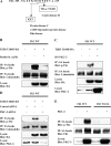
TGF-β mediates Fli1 phosphorylation at a threonine residue(s) through a Smad-independent pathway. 293T cells were transfected with pCTAP-Fli1 along with TβRI T204D, TβRI T204D mL45, or empty vector and incubated for 48 h. Equal amounts of protein from each whole-cell extract were subjected to immunoprecipitation (IP) using streptavidin-coupled agarose beads (SA beads), followed by immunoblotting using antiphosphoserine antibody (p-Ser) or antiphosphothreonine antibody (p-Thr). In order to visualize the total levels of ectopically expressed Fli1, the same membrane was stripped and reprobed with the anti-calmodulin binding peptide antibody. The levels of HA-tagged proteins in cell lysates were determined by Western blotting. To confirm the activation status of TGF-β signaling, the levels of phospho-Smad2 were evaluated by Western blotting. WT, wild type.
FIG. 2.
FIG. 2.
The TGF-β-dependent phosphorylation site(s) of Fli1 is located within the EBD. (A) Schematic representations of pCTAP-Fli1 and three distinct sequential deletion constructs. A major acetylation site, lysine 380, is indicated by an arrow. ATA, A-terminal activation domain. (B) Phosphorylation levels of each Fli1 deletion construct were determined as described in the Fig. 1 legend.
FIG. 3.
FIG. 3.
Fli1 is phosphorylated at threonine 312 in response to TGF-β stimulation. (A) Phosphorylation levels of Fli1 mutants carrying alanine substitutions for threonines 301, 305, 312, and 349 were determined in 293T cells as described in the legend to Fig. 1. (B) Amino acid sequence of a peptide containing phosphorylated threonine, which was used to generate the antiphosphorylation of threonine 312 Fli1 antibody. (C) Determination of the specificity of the phospho-Fli1 (Thr312) antiserum. The blots were prepared as described in the legend to Fig. 1. Strips of the membrane were incubated with phospho-Fli1 (Thr312) antiserum with no additions (left panel), in the presence of 10 μM nonphosphopeptide (non-P-pep) (middle panel) or in the presence of 10 μM nonphosphopeptide and phosphopeptide (P-pep) (right panel). Blots were reprobed with anti-calmodulin binding peptide antibody.
FIG. 4.
FIG. 4.
Fli1 is phosphorylated at threonine 312 through a PKC δ-dependent pathway in response to TGF-β stimulation. (A) The consensus target sites including threonine 312 for serine/threonine kinases. cAMP, cyclic AMP. (B to D) 293T cells were transfected with wild-type pCTAP-Fli1 along with TβRI T204D or empty vector and incubated for 48 h. In some experiments, the cells were treated with 1 μM of rottlerin (B) or transduced with dominant-negative PKC δ-expressing adenovirus (C) for the last 24 h. In the gene silencing experiments, the cells were treated with 10 nM control or PKC δ siRNA using HiPerFect reagent for 24 h prior to plasmid transfection (D). Phosphothreonine levels of Fli1 in each cell lysate were determined by immunoprecipitation (IP) using streptavidin-coupled agarose beads (SA beads), followed by immunoblotting using antiphosphothreonine antibody (p-Thr). The total levels of ectopically expressed Fli1 were determined on the same membrane using anti-calmodulin binding peptide antibody. (E) 293T cells were transfected with expression vectors encoding wild-type Fli1 or the Fli1 T312A mutant and incubated for 48 h. Some cells were transduced with wild-type PKC δ adenovirus for the last 24 h. The levels of Fli1 phosphorylation were determined as described above.
FIG. 5.
FIG. 5.
PKC δ directly phosphorylates Fli1 at threonine 312. (A) Cultured dermal fibroblasts and human microvascular endothelial cells were stained with anti-Fli1 antibody. Positive signals were developed with diaminobenzidine. (B) 293T cells were transfected with TβRI T204D or empty vector for 48 h and then cytoplasmic extracts (500 μl of buffer A) and nuclear extracts (500 μl of buffer C) were prepared. In order to evaluate the ratio of PKC δ localized in the cytoplasm (C) and nucleus (N) accurately, a 10% volume of each extract was subjected to immunoblotting with anti-PKC δ antibody. To confirm that the cytoplasm and nucleus were properly separated, the levels of β-actin and lamin A/C were determined. The bottom panel shows the relative expression levels of PKC δ in each compartment. (C) 293T cells were transfected with the indicated expression vectors for 48 h. Nuclear extracts and whole-cell lysates were prepared under the same conditions. Tagged-Fli1 was precipitated from nuclear extracts with streptavidin-coupled agarose beads (SA beads), and the precipitates were subjected to immunoblotting using anti-PKC δ antibody and anti-calmodulin binding peptide antibody. The levels of PKC δ in the nuclear extracts and the levels of TβRI T204D in the whole-cell lysates were determined by immunoblotting. WT, wild type. (D) Nuclear extracts (NE) and whole-cell lysates were prepared under the same conditions. Nuclear extracts were subjected to DNA affinity precipitation (DNAP) with COL1A2 EBS oligonucleotide. The levels of PKC δ in the precipitates were determined by immunoblotting. (E) Streptavidin bead-bound tagged Fli1 prepared from 293T cells was incubated with recombinant PKC δ-GST fusion protein. In order to activate PKC δ, phosphatidyl serine and phorbol ester were added to the reaction. The whole reaction was subjected to immunoblotting using anti-phospho-Fli1 (Thr312) antibody. The levels of tagged Fli1 and PKC δ were confirmed by immunoblotting on the same membrane.
FIG. 6.
FIG. 6.
Phosphorylation at threonine 312 increases the interaction of Fli1 with PCAF and subsequently promotes its acetylation. (A) Wild-type Fli1 or Fli1-T312A constructs were transfected into 293T cells along with the indicated expression vectors, including TβRI T204D, PCAF, and PCAF/ΔHAT, and incubated for 48 h. Total cell extracts were subjected to immunoprecipitation (IP) using streptavidin-coupled agarose beads (SA beads), followed by immunoblotting using anti-acetylated lysine antibody (AcK) or anti-Flag antibody. In order to visualize the total levels of ectopically expressed Fli1, the same membrane was stripped and reprobed with the anti-calmodulin binding peptide antibody. The levels of Flag-tagged and HA-tagged proteins in cell lysates were determined by Western blotting. (B) Wild-type Fli1 or Fli1-T312A constructs were transfected into 293T cells along with PCAF or PCAF/ΔHAT, and the cells were incubated for 48 h. The DNA binding ability of each Fli1 construct was evaluated by DNA affinity precipitation (DNAP) assay. The levels of Fli1 and Flag-tagged proteins were determined by immunoblotting. (C) 293T cells were transfected with the −772 human COL1A2 promoter, along with the indicated expression vectors. After 48 h incubation, formaldehyde-cross-linked, moderately sheared chromatin was prepared. The DNA fragments were immunoprecipitated using rabbit polyclonal anti-Fli1 antibody, and the presence of COL1A2 promoter fragments were detected using PCR.
FIG. 7.
FIG. 7.
The phosphorylation of Fli1 precedes its acetylation in dermal fibroblasts in response to TGF-β. (A) Subconfluent dermal fibroblasts were serum starved for 24 h and then treated with TGF-β1 (2.5 ng/ml) for the indicated periods of time. Fli1 was precipitated from nuclear extracts using mouse monoclonal anti-Fli1 antibody, and the phosphorylation levels of Fli1 at threonine 312 were determined by immunoblotting with anti-phospho-Fli1 (Thr312) antibody. The membrane was stripped and reprobed with rabbit polyclonal anti-Fli1 antibody. The graph shows the levels of phosphorylation normalized by total Fli1 levels. (B) Subconfluent dermal fibroblasts were serum starved for 24 h and then treated with TGF-β1 (2.5 ng/ml) for the indicated periods of time. The phosphorylation and acetylation levels of Fli1 were visualized on the same membrane by sequential Western blots. (C) Fli1 was precipitated from nuclear extracts using monoclonal mouse anti-Fli1 antibody, and the precipitates were subjected to immunoblotting with anti-PKC δ antibody. The membrane was stripped and reprobed with rabbit polyclonal anti-Fli1 antibody. (D) Subconfluent dermal fibroblasts were serum starved for 48 h. Where indicated, for the last 24 h, cells were treated with rottlerin (3 μM), anacardic acid (10 μM), or a corresponding volume of dimethyl sulfoxide. The levels of Fli1 phosphorylation and acetylation were determined as described above. AcK, anti-acetylated lysine antibody. (E and F) Dermal fibroblasts were transduced with PCAF siRNA adenovirus (with scrambled siRNA adenovirus as a control) or transfected with PKC δ siRNA oligonucleotide (with control siRNA oligonucleotide as a control) using HiPerFect reagent for 72 h. Where indicated, cells were treated with TGF-β for the last 3 h. The levels of Fli1 phosphorylation and acetylation were determined as described above. (G and H) Dermal fibroblasts were incubated for 3 h with TGF-β (2.5 ng/ml) or a vehicle in the presence (+) or absence (−) of rottlerin (3 μM). For the gene silencing experiments, cells were treated with PKC δ siRNA or control siRNA for 72 h. Fli1 was precipitated from nuclear extracts using monoclonal mouse anti-Fli1 antibody, and the precipitates were subjected to immunoblotting with anti-PCAF antibody. The membrane was stripped and reprobed with rabbit polyclonal anti-Fli1 antibody. (I and J) Dermal fibroblasts were treated overnight with rottlerin (3 μM) or dimethyl sulfoxide (DMSO). For the gene silencing experiments, cells were treated with PKC δ siRNA or control siRNA for 72 h. The levels of Fli1 phosphorylation and acetylation were determined as described above. (K) Dermal fibroblasts were treated with PKC δ siRNA or control siRNA for 72 h. In some experiments, cells were stimulated with TGF-β1 (2.5 ng/ml) for the last 3 h. Formaldehyde-cross-linked, moderately sheared chromatin was prepared. The DNA fragments were immunoprecipitated using rabbit anti-Fli1 antibody (Anti-Fli1), and the presence of the human COL1A2 promoter fragments were detected using PCR. No-Ab, beads alone.
FIG. 8.
FIG. 8.
Phosphorylation at threonine 312 is essential for the regulation of the transcriptional activity of Fli1 in the context of the human COL1A2 promoter. (A and B) Dermal fibroblasts were transduced with PKC δ-expressing adenovirus (with green fluorescent protein-expressing adenovirus as a control) or transfected with PKC δ siRNA oligonucleotide (with control siRNA oligonucleotide as a control) for 72 h. The protein levels of type I collagen, PKC δ, and β-actin were determined by immunoblotting using whole-cell extracts. mRNA levels of COL1A2 gene were determined by quantitative real-time PCR under the same conditions. (C) Fibroblasts were transfected with the −772 COL1A2/CAT construct (2 μg), along with Fli1 constructs (0.1 μg), either the wild-type or the T312A mutant, and either PCAF or empty vector (0.5 μg) for 48 h. For the last 24 h, some cells were stimulated with TGF-β1 (2.5 ng/ml). Values represent CAT activities relative to those of untreated cells (100 arbitrary units [AU]). The mean and SD of the results from three separate experiments are shown. *, P < 0.05 versus control cells stimulated with TGF-β1; **, P < 0.05 versus cells transfected with Fli1 only and stimulated with TGF-β1. (D) Fibroblasts were transfected with 2 μg of the −772 COL1A2/CAT construct, along with empty vector or the indicated Fli1 constructs, and incubated for 48 h. At 6 h after transfection, some cells were transduced with PKC δ-expressing adenovirus (at multiplicities of infection of 5 and 10). In addition, some cells were treated with TGF-β1 (2.5 ng/ml) for the last 24 h. Values represent CAT activities relative to those of untreated cells transfected with empty vector (100 AU). The means and SD of the results from three separate experiments are shown. *, P < 0.05 versus control cells stimulated with TGF-β1; **, P < 0.05 versus cells transfected with wild-type Fli1 in the absence of PKC δ-expressing adenovirus and stimulated with TGF-β1; #, P < 0.05 versus cells transfected with wild-type Fli1 in the presence of PKC δ-expressing adenovirus and stimulated with TGF-β1.
FIG. 9.
FIG. 9.
Rottlerin increases the binding of Fli1 to the COL1A1 promoter. (A) Chromatin was isolated from adult dermal fibroblasts treated with PKC δ siRNA or control siRNA and immunoprecipitated using rabbit anti-Fli1 antibody (5 μg) or beads alone (No-Ab). After the isolation of bound DNA, PCR amplification was carried out using the COL1A1 promoter-specific primers. Input DNA was taken from each sample before the addition of an antibody. (B) Confluent fibroblasts were serum starved for 48 h. Some cells were treated with rottlerin (3 μM) for the last 24 h. Nuclear extracts were incubated with biotin-labeled oligonucleotides. Proteins bound to these nucleotides were isolated with streptavidin-coupled agarose beads, and Fli1 was detected by immunoblotting. The levels of Fli1 in nuclear extracts were determined by Western blotting. WT, wild type; DNAP, DNA affinity precipitation.
FIG. 10.
FIG. 10.
Schematic model for the TGF-β-induced posttranslational modifications of Fli1. In quiescent cells, collagen gene expression is repressed by Fli1 occupancy of the collagen promoter (1). In response to TGF-β stimulation, PKC δ is translocated into the nucleus and recruited to the COL1A2 promoter, leading to the phosphorylation of Fli1 at threonine 312. The phosphorylation of Fli1 is required for the subsequent PCAF-mediated acetylation of Fli1 at lysine 380. Acetylated Fli1 dissociates from the collagen promoter, resulting in the enhancement of the collagen gene transcription.

References

    1. Asano, Y., J. Czuwara, and M. Trojanowska. 2007. Transforming growth factor-beta regulates DNA binding activity of transcription factor Fli1 by p300/CREB-binding protein-associated factor-dependent acetylation. J. Biol. Chem. 28234672-34683. - PubMed
    1. Asano, Y., H. Ihn, K. Yamane, M. Jinnin, Y. Mimura, and K. Tamaki. 2004. Phosphatidylinositol 3-kinase is involved in alpha2(I) collagen gene expression in normal and scleroderma fibroblasts. J. Immunol. 1727123-7135. - PubMed
    1. Asano, Y., H. Ihn, K. Yamane, M. Kubo, and K. Tamaki. 2004. Impaired Smad7-Smurf-mediated negative regulation of TGF-beta signaling in scleroderma fibroblasts. J. Clin. Investig. 113253-264. - PMC - PubMed
    1. Asano, Y., M. Markiewicz, M. Kubo, G. Szalai, D. K. Watson, and M. Trojanowska. 2009. Transcription factor Fli1 regulates collagen fibrillogenesis in mouse skin. Mol. Cell. Biol. 29425-434. - PMC - PubMed
    1. Bailly, R.-A., R. Bosselut, J. Zucman, F. Cormier, O. Delattre, M. Roussel, G. Thomas, and J. Ghysdael. 1994. DNA-binding and transcriptional activation properties of the EWS-FLI-1 fusion protein resulting from the t(11;22) translocation in Ewing sarcoma. Mol. Cell. Biol. 143230-3241. - PMC - PubMed

Publication types

MeSH terms