. 2009 Jan 21;29(3):678-85.
doi: 10.1523/JNEUROSCI.5060-08.2009.

Endogenous truncated TrkB.T1 receptor regulates neuronal complexity and TrkB kinase receptor function in vivo

Affiliations

Endogenous truncated TrkB.T1 receptor regulates neuronal complexity and TrkB kinase receptor function in vivo

Laura Carim-Todd et al. J Neurosci. .

Abstract

Pathological or in vitro overexpression of the truncated TrkB (TrkB.T1) receptor inhibits signaling through the full-length TrkB (TrkB.FL) tyrosine kinase receptor. However, to date, the role of endogenous TrkB.T1 is still unknown. By studying mice lacking the truncated TrkB.T1 isoform but retaining normal spatiotemporal expression of TrkB.FL, we have analyzed TrkB.T1-specific physiological functions and its effect on endogenous TrkB kinase signaling in vivo. We found that TrkB.T1-deficient mice develop normally but show increased anxiety in association with morphological abnormalities in the length and complexity of neurites of neurons in the basolateral amygdala. However, no behavioral abnormalities were detected in hippocampal-dependent memory tasks, which correlated with lack of any obvious hippocampal morphological deficits or alterations in basal synaptic transmission and long-term potentiation. In vivo reduction of TrkB signaling by removal of one BDNF allele could be partially rescued by TrkB.T1 deletion, which was revealed by an amelioration of the enhanced aggression and weight gain associated with BDNF haploinsufficiency. Our results suggest that, at the physiological level, TrkB.T1 receptors are important regulators of TrkB.FL signaling in vivo. Moreover, TrkB.T1 selectively affects dendrite complexity of certain neuronal populations.


Figures
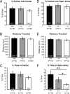
Figure 1.
Figure 1.
TrkB.T1-deficient animals have increased anxiety-like behavior. The behavior of +/+, +/−, and −/− TrkB.T1 mice was analyzed using the open-field (A–C) and the elevated-plus maze (D–F) tests. Mouse performances were video recorded and analyzed blindly. For the open-field test, the number of mouse entries into the center (A), distance traveled (B), and time spent in the center of the arena (C) were determined. Anxiety level was measured by the relative amount of exploration devoted to the central quadrants relative to those located adjacent to the walls of the arena. For the elevated-plus maze test, the number of entries into the open arms (D), distance traveled (E), and time spent in the open arms (F) were scored. Anxiety was measured by the relative amount of exploration invested in the open arms relative to that of the enclosed arms. For each test, mouse behavior was followed for 10 min. *p < 0.05; **p < 0.01.
Figure 2.
Figure 2.
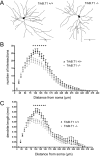
TrkB.T1−/− mice have decreased neuronal complexity in the amygdala. Morphological analysis of basolateral amygdala neurons visualized by the rapid Golgi impregnation method. A, Neurolucida representation of a typical stained basolateral amygdala neuron from TrkB.T1−/− (right) and wild-type control (left) mouse brain sections. Sholl analysis of the number of dendrite intersections (B) and length of dendrites of amygdala neurons (C). Postnatal day 60 mice and 25 neurons per mouse were used in the analysis. All results are presented as means ± SEM determined from analyzing six mice per genotype. *p < 0.01.
Figure 3.
Figure 3.
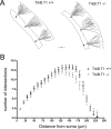
Loss of TrkB.T1 does not affect hippocampus neurite morphology. A, Representative examples of Golgi-stained dentate gyrus neurons from postnatal day 60 wild-type (+/+; left) and TrkB.T1-deficient (−/−; right) mice. Sholl analyses of the number of intersections (B) in hippocampal dentate gyrus neurons. All results are presented as means ± SEM determined by analysis of six mice per genotype and 25 neurons per mouse. *p < 0.01.
Figure 4.
Figure 4.
TrkB.T1 does not affect hippocampal basal synaptic transmission and LTP. A, Input–output curve: initial slope of fEPSPs obtained with increased stimulus intensity were plotted versus the response normalized to the maximal response obtained (n = 5 per genotype). B, fEPSP recorded in wild-type and mutant TrkB.T1 mice. The average of five traces obtained before (gray) and 1 h after (black) the LTP conditioning protocol (2 times at 250 ms, 100 Hz train) are represented. C, Time course of initial slope of fEPSP before and after LTP induction in wild-type (black diamonds; n = 12) and TrkB.T1−/− (gray squares; n = 11) mice.
Figure 5.
Figure 5.
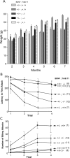
TrkB.T1 deletion partially rescues the obesity and the aggressive phenotype caused by partial loss of BDNF. A, Weight analysis of wild-type, TrkB.T1−/−, TrkB.T1−/−;BDNF+/−, TrkB.T1+/−;BDNF+/−, and BDNF+/− mice. Mice had ad libitum access to food and water. The weight of the indicated mouse groups was monitored once a month until mice reached an age of 7 months. Bars indicate means ± SEM. Each group comprised a minimum of eight mice. At 4 months of age, one-way ANOVA statistical analysis followed by Tukey's multiple comparison test shows that the weight of BDNF+/−;TrkB.T1−/− is significantly different from that of BDNF+/− (*p < 0.05) mice but not from that of wild-type animals. At 5, 6, and 7 months, the weights of BDNF+/−;TrkB.T1−/− are significantly different from both wild-type and BDNF+/− mice (*p < 0.05). B, C, Aggressive behavior was assessed for each of the above-indicated genotype using the resident/intruder paradigm. The latency to first attack (B) was measured on 4 consecutive days (1 session of 5 min a day) for each genotype using an age- and weight-matched wild-type intruder mouse. The number of mice for each group is indicated in parentheses. Means ± SEM bars are indicated. Latency to first attack in trial 3 as analyzed by one-way ANOVA and Tukey's multiple comparison test reveals a significant difference between BDNF+/− and BDNF+/−;TrkB.T1+/− (p < 0.05) but not between BDNF+/+;TrkB.T1+/+ and BDNF+/−;TrkB.T1+/−. C, Number of biting attacks over a period of 5 min. The number of biting attacks by BDNF+/− mice in trials 2–4 is significantly different (p < 0.05) from that of BDNF+/−;TrkB.T1+/− but not of BDNF+/−;TrkB.T1−/− mice.

References

    1. Allendoerfer KL, Cabelli RJ, Escandón E, Kaplan DR, Nikolics K, Shatz CJ. Regulation of neurotrophin receptors during the maturation of the mammalian visual system. J Neurosci. 1994;14:1795–1811. - PMC - PubMed
    1. Baxter GT, Radeke MJ, Kuo RC, Makrides V, Hinkle B, Hoang R, Medina-Selby A, Coit D, Valenzuela P, Feinstein SC. Signal transduction mediated by the truncated trkB receptor isoforms, trkB.T1 and trkB.T2. J Neurosci. 1997;17:2683–2690. - PMC - PubMed
    1. Bibel M, Barde YA. Neurotrophins: key regulators of cell fate and cell shape in the vertebrate nervous system. Genes Dev. 2000;14:2919–2937. - PubMed
    1. Biffo S, Offenhäuser N, Carter BD, Barde YA. Selective binding and internalisation by truncated receptors restrict the availability of BDNF during development. Development. 1995;121:2461–2470. - PubMed
    1. Bothwell M. Functional interactions of neurotrophins and neurotrophin receptors. Annu Rev Neurosci. 1995;18:223–253. - PubMed

Publication types

MeSH terms

Substances

LinkOut - more resources