. 2009 Jan 21;29(3):842-51.
doi: 10.1523/JNEUROSCI.4434-08.2009.

Impaired synaptic vesicle release and immaturity of neuromuscular junctions in spinal muscular atrophy mice

Affiliations

Impaired synaptic vesicle release and immaturity of neuromuscular junctions in spinal muscular atrophy mice

Lingling Kong et al. J Neurosci. .

Abstract

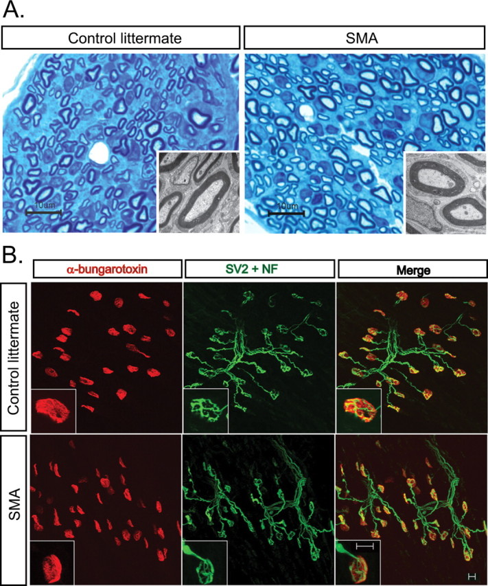
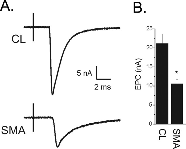
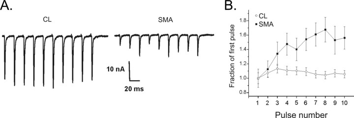
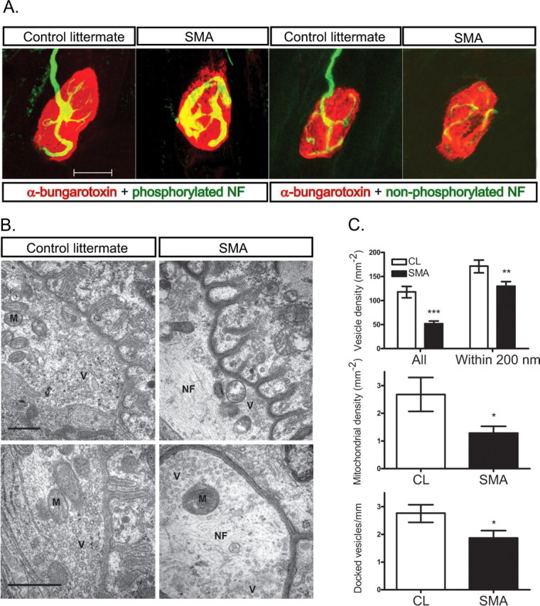
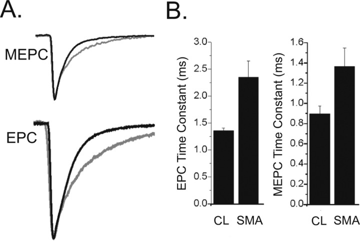
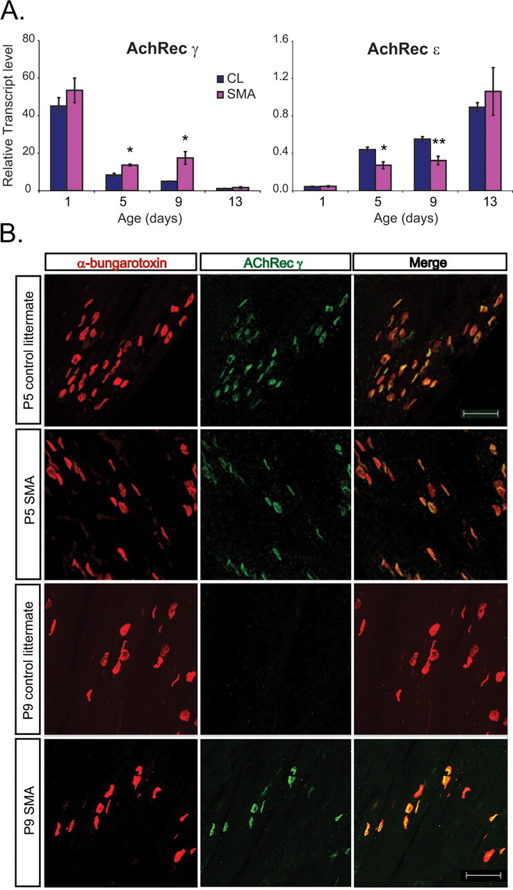
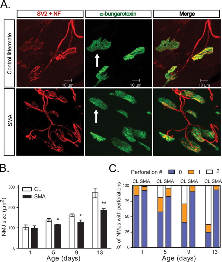
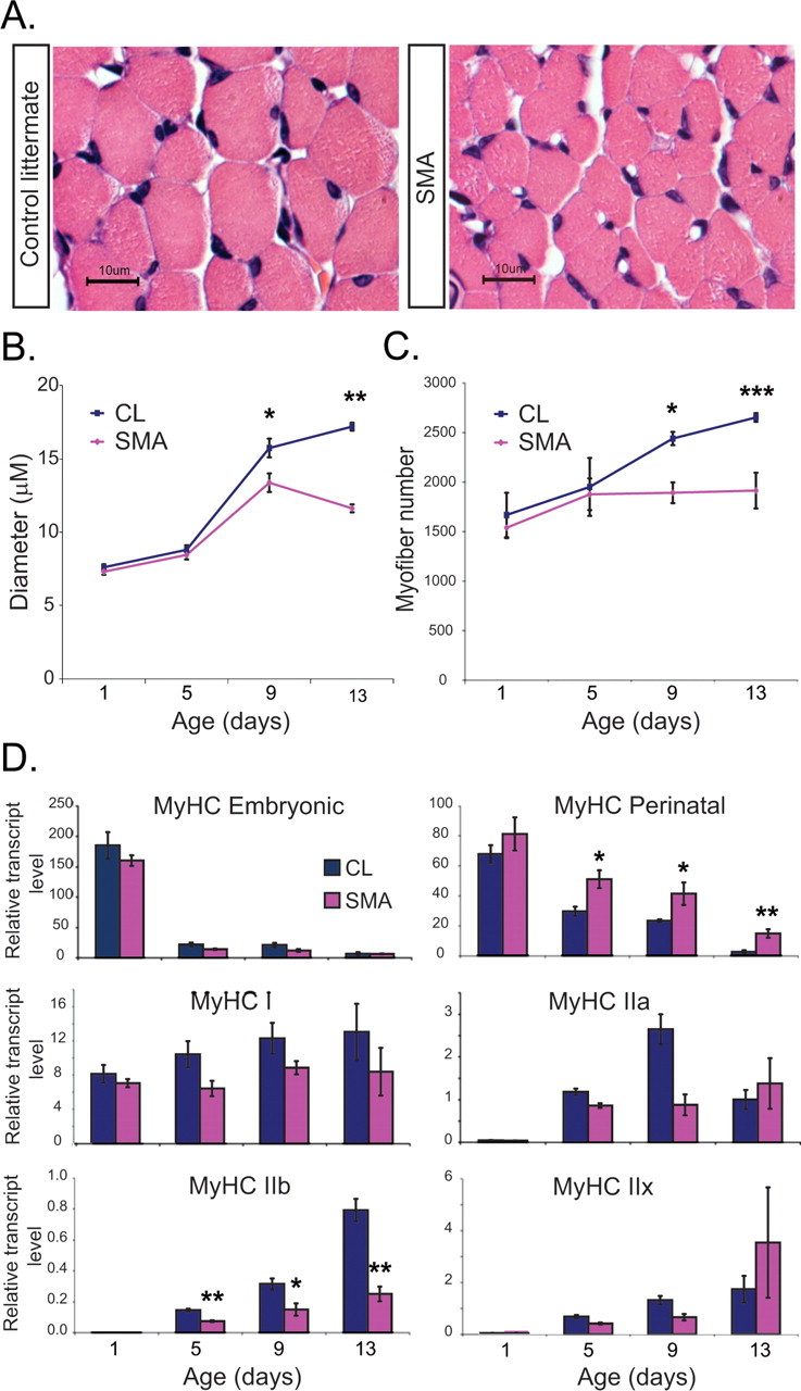
The motor neuron disease spinal muscular atrophy (SMA) causes profound muscle weakness that most often leads to early death. At autopsy, SMA is characterized by loss of motor neurons and muscle atrophy, but the initial cellular events that precipitate motor unit dysfunction and loss remain poorly characterized. Here, we examined the function and corresponding structure of neuromuscular junction (NMJ) synapses in a mouse model of severe SMA (hSMN2/delta7SMN/mSmn-/-). Surprisingly, most SMA NMJs remained innervated even late in the disease course; however they showed abnormal synaptic transmission. There was a two-fold reduction in the amplitudes of the evoked endplate currents (EPCs), but normal spontaneous miniature EPC (MEPC) amplitudes. These features in combination indicate reduced quantal content. SMA NMJs also demonstrated increased facilitation suggesting a reduced probability of vesicle release. By electron microscopy, we found a decreased density of synaptic vesicles that is likely to contribute to the reduced release probability. In addition to presynaptic defects, there were postsynaptic abnormalities. EPC and MEPC decay time constants were prolonged because of a slowed switch from the fetal acetylcholine receptor (AChR) gamma-subunit to the adult epsilon-subunit. There was also reduced size of AChR clusters and small myofibers, which expressed an immature pattern of myosin heavy chains. Together these results indicate that impaired synaptic vesicle release at NMJs in severe SMA is likely to contribute to failed postnatal maturation of motor units and muscle weakness.


Figures

Figure 1.
Figure 1.
Limited denervation of SMA NMJs. A, Intramuscular axons examined by light microscopy and EM in the paraspinal muscle of P13 SMA and CL mice show normal axon and myelin sheath structure and no axonal degeneration. Scale bar, 10 μm and inset EM. B, IHC for NMJs in P13 TA muscle of SMA and CL mice showing that NMJs are well innervated. Scale bar, 10 μm.
Figure 2.
Figure 2.
Decreased EPC amplitude in SMA NMJs. A, Shown are representative EPCs from a CL and a SMA NMJ. The EPC from the SMA NMJ is less than ½ the amplitude of the CL. The stimulus artifact from nerve stimulation precedes the NMJ current by several milliseconds (ms). B, Shown is a bar graph of the average EPC amplitude from CL and SMA mice (*p < 0.01, n = 5 CL and 6 SMA mice).
Figure 3.
Figure 3.
Reduction in quantal content underlies the reduction in EPC amplitude in SMA mice. A, Shown are representative average MEPCs from a CL and a SMA NMJ. There is no difference in amplitude. B, Shown are bar graphs of the average MEPC amplitude from CL and SMA pups (p = 0.38) and of quantal content (*p < 0.01).
Figure 4.
Figure 4.
Facilitation is increased in SMA NMJs. A, Shown are EPCs during a 50 Hz train of pulses for CL and SMA NMJs. In the CL NMJ there is a slight increase in EPC amplitude early in the train. By the end of the train, however, EPC amplitude has decreased back to the initial value. In the SMA NMJ, the initial EPC of the train is small, but there is an increase in EPC amplitude during the train. In both the CL and the SMA trace, stimulus artifacts were removed for clarity. B, The normalized change in average EPC amplitudes is plotted during trains of 50 Hz pulses for CL (n = 35 endplates from 3 muscles) and SMA NMJs (n = 17 endplates from 2 muscles). By the third pulse of the train, the SMA NMJs have facilitated to a greater degree that CL endplates. The facilitation is sustained throughout the train such that it is still statistically significant on the 10th pulse (p < 0.05).
Figure 5.
Figure 5.
Decreased synaptic vesicle density at SMA NMJs. A, IHC for phosphorylated NFs and nonphosphorylated NFs shows accumulation of phosphorylated NFs within SMA presynaptic terminals. B, Electron microphotographs of NMJs in P13 TA muscle isolated from CL and SMA mice. Synaptic vesicles (V) are more homogeneously and densely distributed in CL compared with SMA mice. Accumulations of NF (N) are evident particularly at the center of SMA presynaptic terminals. Mitochondria (M) are reduced in number, but have normal morphology in SMA mice. Scale bar, 0.5 μm. C, Quantification of total synaptic vesicle density, synaptic density within 200 nm of the membrane, docked vesicle number, and mitochondria density (*p < 0.05, **p < 0.01, ***p < 0.001).
Figure 6.
Figure 6.
EPC and MEPC decay time constants are prolonged in NMJs from SMA mice. A, The CL and SMA MEPCs and EPCs from Figures 2 and 3 have been normalized and aligned by their peaks to allow for comparison of rates of decay. The MEPC and EPC from the SMA pups are gray and have slower rates of decay. B, Shown are bar graphs of the average EPC decay (*p = 0.02) and MEPC decay (p = 0.06) in CL and SMA NMJs.
Figure 7.
Figure 7.
Delayed switch of AChR subunits in SMA NMJs. A, AChR γ and AChR ε transcript levels in all hindlimb muscle in SMA and CL mice at P1, P5, P9, and P13 show increased expression of AChR γ and reduced expression of AChR ε at P5 and P9 (*p < 0.05, **p < 0.005). B, IHC for AChR γ in the TA muscle indicates persistent expression at P9 SMA NMJs when expression is no longer evident in CL mice.
Figure 8.
Figure 8.
Simplified morphology of SMA NMJs. A, NMJs in paraspinal muscle of P13 SMA and CL mice showing simplification of the morphology of the postsynaptic terminals. Scale bar, 10 μm. B, NMJ area is decreased in the TA muscle of SMA mice at P5, P9, and P13 (*p < 0.05, **p < 0.005). C, SMA NMJs in the TA muscle have decreased numbers of perforations. The number of NMJs with no perforations is increased in SMA mice at P5, P9 (p < 0.05), and P13 (p < 0.005).
Figure 9.
Figure 9.
Impaired SMA neonatal muscle growth. A, Cross-sections of TA muscle stained with H&E from P13 CL and SMA mice showing reduced myofiber diameter in SMA mice. B, Quantification of average myofiber diameter in the TA muscle at P1, P5, P9, and P13 (n = 3 CL and 3 SMA mice per time point). C, Quantification of total myofiber number in TA muscle at P1, P5, P9, and P13 (n = 3 CL and 3 SMA mice per time point). D, Embryonic MyHC, perinatal MyHC, MyHC type I, MyHC type IIa, MyHC IIb, and MyHC type IIx transcript levels were quantified in all hind-limb muscle in SMA and CL mice at P1, P5, P9, and P13. Perinatal MyHC levels are increased and adult MyHC isoforms are reduced in SMA mice compared with CLs starting at P5 (n = 4–6 CL and SMA mice per time point, *p < 0.05, **p < 0.01, ***p < 0.005).

References

    1. Avila AM, Burnett BG, Taye AA, Gabanella F, Knight MA, Hartenstein P, Cizman Z, Di Prospero NA, Pellizzoni L, Fischbeck KH, Sumner CJ. Trichostatin A increases SMN expression and survival in a mouse model of spinal muscular atrophy. J Clin Invest. 2007;117:659–671. - PMC - PubMed
    1. Balice-Gordon RJ, Smith DB, Goldman J, Cork LC, Shirley A, Cope TC, Pinter MJ. Functional motor unit failure precedes neuromuscular degeneration in canine motor neuron disease. Ann Neurol. 2000;47:596–605. - PubMed
    1. Braun S, Croizat B, Lagrange MC, Warter JM, Poindron P. Constitutive muscular abnormalities in culture in spinal muscular atrophy. Lancet. 1995;345:694–695. - PubMed
    1. Chang HC, Dimlich DN, Yokokura T, Mukherjee A, Kankel MW, Sen A, Sridhar V, Fulga TA, Hart AC, Van Vactor D, Artavanis-Tsakonas S. Modeling spinal muscular atrophy in Drosophila. PLoS ONE. 2008;3:e3209. - PMC - PubMed
    1. Cifuentes-Diaz C, Nicole S, Velasco ME, Borra-Cebrian C, Panozzo C, Frugier T, Millet G, Roblot N, Joshi V, Melki J. Neurofilament accumulation at the motor endplate and lack of axonal sprouting in a spinal muscular atrophy mouse model. Hum Mol Genet. 2002;11:1439–1447. - PubMed

Publication types

MeSH terms

Substances