. 2009 Apr;296(4):F859-66.
doi: 10.1152/ajprenal.90238.2008. Epub 2009 Jan 21.

SGK1-sensitive renal tubular glucose reabsorption in diabetes

Affiliations

SGK1-sensitive renal tubular glucose reabsorption in diabetes

Teresa F Ackermann et al. Am J Physiol Renal Physiol. 2009 Apr.

Abstract

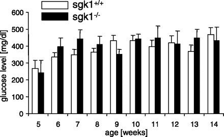
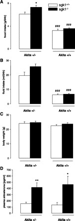
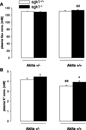
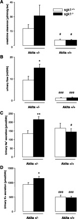
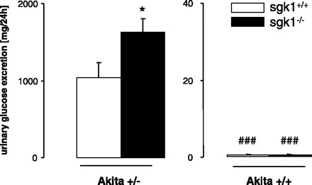
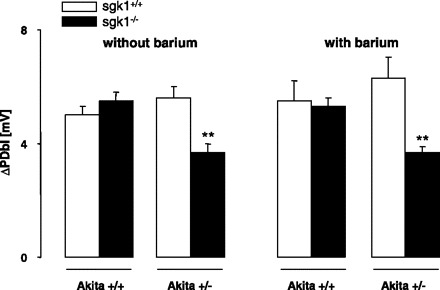
The hyperglycemia of diabetes mellitus increases the filtered glucose load beyond the maximal tubular transport rate and thus leads to glucosuria. Sustained hyperglycemia, however, may gradually increase the maximal renal tubular transport rate and thereby blunt the increase of urinary glucose excretion. The mechanisms accounting for the increase of renal tubular glucose transport have remained ill-defined. A candidate is the serum- and glucocorticoid-inducible kinase SGK1. The kinase has been shown to stimulate Na(+)-coupled glucose transport in vitro and mediate the stimulation of electrogenic intestinal glucose transport by glucocorticoids in vivo. SGK1 expression is confined to glomerula and distal nephron in intact kidneys but may extend to the proximal tubule in diabetic nephropathy. To explore whether SGK1 modifies glucose transport in diabetic kidneys, Akita mice (akita(+/-)), which develop spontaneous diabetes, have been crossbred with gene-targeted mice lacking SGK1 on one allele (sgk1(+/-)) to eventually generate either akita(+/-)/sgk1(-/-) or akita(+/-)/sgk1(+/+) mice. Both akita(+/-)/sgk1(-/-) and akita(+/-)/sgk1(+/+) mice developed profound hyperglycemia (>20 mM) within approximately 6 wk. Body weight and plasma glucose concentrations were not significantly different between these two genotypes. However, urinary excretion of glucose and urinary excretion of fluid, Na(+), and K(+), as well as plasma aldosterone concentrations, were significantly higher in akita(+/-)/sgk1(-/-) than in akita(+/-)/sgk1(+/+) mice. Studies in isolated perfused proximal tubules revealed that the electrogenic glucose transport was significantly lower in akita(+/-)/sgk1(-/-) than in akita(+/-)/sgk1(+/+) mice. The data provide the first evidence that SGK1 participates in the stimulation of renal tubular glucose transport in diabetic kidneys.


Figures

Fig. 1.
Fig. 1.
Plasma glucose concentrations in akita+/−/sgk1−/− and akita+/−/sgk1+/+ mice. Values are means ± SE (for akita+/−/sgk1−/− mice/akita+/−/sgk1+/+ mice, n = 3/5, 7/8, 9/11, 9/11, 7/11, 8/9, 6/11, 6/7, 9/9, and 7/10 for weeks 5–14, respectively) of plasma glucose concentrations in fed mice carrying the Akita mutation (akita+/−) and either lacking (sgk1−/−) or expressing (sgk1+/+) functional SGK1. Blood glucose concentrations were determined regularly between ages 5 and 14 wk.
Fig. 2.
Fig. 2.
Food and fluid intake, body weight, and plasma aldosterone concentrations in diabetic akita+/−/sgk1−/− and akita+/−/sgk1+/+ mice and in nondiabetic akita+/+/sgk1−/− and akita+/+/sgk1+/+ mice. Values are means ± SE (n = 7–10 for each group) of food intake (A), fluid intake (B), body weight (C), and plasma aldosterone concentrations (D) in mice carrying the Akita mutation (akita+/−) and either lacking (sgk1−/−) or expressing (sgk1+/+) functional SGK1, as well as in nondiabetic mice (akita+/+) either lacking or expressing functional SGK1. *P < 0.05 vs. respective value of akita+/−/sgk1+/+ mice. ###P < 0.001 vs. respective value of akita+/− mice.
Fig. 3.
Fig. 3.
Plasma Na+ and K+ concentrations in diabetic akita+/−/sgk1−/− and akita+/−/sgk1+/+ mice and in nondiabetic akita+/+/sgk1−/− and akita+/+/sgk1+/+ mice. Values are means ± SE (n = 6–8 for each group) of plasma Na+ (A) and K+ concentrations (B) in mice carrying the Akita mutation (akita+/−) and either lacking (sgk1−/−) or expressing (sgk1+/+) functional SGK1. Nondiabetic mice (akita+/+) served as controls. *P < 0.05 vs. respective value of akita+/+/sgk1+/+ mice. ##P < 0.01 vs. respective value of akita+/− mice.
Fig. 4.
Fig. 4.
Urinary flow rate, creatinine clearance, and urinary excretion of Na+ and K+ in diabetic akita+/−/sgk1−/− and akita+/−/sgk1+/+ mice and in nondiabetic akita+/+/sgk1−/− and akita+/+/sgk1+/+ mice. Value are means ± SE (n = 5–8 for each group) of urinary flow rate (A), creatinine clearance (B), and urinary excretion of Na+ (C) and K+ (D) in mice carrying the Akita mutation (akita+/−) and either lacking (sgk1−/−) or expressing (sgk1+/+) functional SGK1. Nondiabetic mice (akita+/+) served as controls. *P < 0.05; **P < 0.01 vs. respective value of akita+/−/sgk1+/+ mice. #P < 0.05; ###P < 0.001 vs. respective value of akita+/− mice.
Fig. 5.
Fig. 5.
Urinary glucose excretion in diabetic akita+/−/sgk1−/− and akita+/−/sgk1+/+ mice and in nondiabetic akita+/+/sgk1−/− and akita+/+/sgk1+/+ mice. Values are means ± SE (n = 6–8 for each group) of urinary glucose excretion in mice carrying the Akita mutation (akita+/−) and either lacking (sgk1−/−; average age = 5.54 mo) or expressing (sgk1+/+; average age = 4.26 mo) functional SGK1. Nondiabetic mice (akita+/+; average age 2.83 mo) served as controls. *P < 0.05 vs. respective value of akita+/−/sgk1+/+ mice. ###P < 0.001 vs. respective value of akita+/− mice.
Fig. 6.
Fig. 6.
Glucose-induced depolarization of the basolateral cell membrane in diabetic akita+/−/sgk1−/− and akita+/−/sgk1+/+ mice and in nondiabetic akita+/+/sgk1−/− and akita+/+/sgk1+/+ mice. Values are means ± SE (n = 5–8 for each group) of the depolarization of the basolateral cell membrane (ΔPDbl) by addition of 20 mM glucose (replacing 20 mM mannitol) in isolated perfused proximal tubules from mice carrying the Akita mutation (akita+/−) and either lacking (sgk1−/−) or expressing (sgk1+/+ mice) functional SGK1. Nondiabetic mice (akita+/+) served as controls. **P < 0.01 vs. respective value of akita+/−/sgk1+/+ mice.
Fig. 7.
Fig. 7.
In situ hybridization for SGK1 mRNA on kidney sections of normal and diabetic mice. A and B: overview of SGK1 mRNA expression in the kidney of normal (A) and diabetic mice (B). Arrows point to the strong signals in the collecting duct epithelium of the renal papilla. C: overview of diabetic kidney section hybridized with the sense probe. D and E: higher magnification of the renal cortex of normal (D) and diabetic mice (E). F: higher magnification of the renal cortex of diabetic kidney section hybridized with the sense probe. rc, Renal cortex; rm, renal medulla. Scale bars: A–C, 500 μm; D–F, 20 μm.
Fig. 8.
Fig. 8.
Combined in situ hybridization for SGK1 mRNA and SGLT1 immunohistochemistry on kidney sections of diabetic mice. A: overview of SGK1 mRNA (violet) and SGLT1 expression (brown) in the renal cortex of diabetic mice. B and C: higher magnification of the renal cortex. Arrows point to double-labeled tubular cells coexpressing SGK1 mRNA (violet) and SGLT1 (brown). Arrowheads indicate SGK-1 mRNA-expressing cells that are negative for SGLT1. Scale bars: A, 50 μm; B and C, 20 μm.

References

    1. Akutsu N, Lin R, Bastien Y, Bestawros A, Enepekides DJ, Black MJ, White JH. Regulation of gene expression by 1alpha,25-dihydroxyvitamin D3 and Its analog EB1089 under growth-inhibitory conditions in squamous carcinoma Cells. Mol Endocrinol 15: 1127–1139, 2001 - PubMed
    1. Alvarez de la Rosa D, Zhang P, Naray-Fejes-Toth A, Fejes-Toth G, Canessa CM. The serum and glucocorticoid kinase sgk increases the abundance of epithelial sodium channels in the plasma membrane of Xenopus oocytes. J Biol Chem 274: 37834–37839, 1999 - PubMed
    1. Alvarez de la Rosa D, Canessa CM. Role of SGK in hormonal regulation of epithelial sodium channel in A6 cells. Am J Physiol Cell Physiol 284: C404–C414, 2003 - PubMed
    1. Belaiba RS, Djordjevic T, Bonello S, Artunc F, Lang F, Hess J, Gorlach A. The serum- and glucocorticoid-inducible kinase Sgk-1 is involved in pulmonary vascular remodeling: role in redox-sensitive regulation of tissue factor by thrombin. Circ Res 98: 828–836, 2006 - PubMed
    1. Bhargava A, Fullerton MJ, Myles K, Purdy TM, Funder JW, Pearce D, Cole TJ. The serum- and glucocorticoid-induced kinase is a physiological mediator of aldosterone action. Endocrinology 142: 1587–1594, 2001 - PubMed

Publication types

MeSH terms