. 2009 Mar;20(6):1816-32.
doi: 10.1091/mbc.e08-09-0894. Epub 2009 Jan 21.

SCAMP3 negatively regulates epidermal growth factor receptor degradation and promotes receptor recycling

Affiliations

SCAMP3 negatively regulates epidermal growth factor receptor degradation and promotes receptor recycling

Quyen L Aoh et al. Mol Biol Cell. 2009 Mar.

Abstract

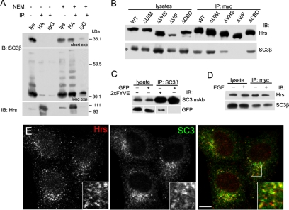
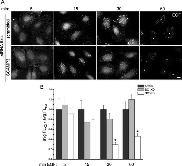
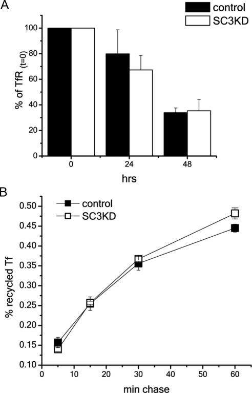
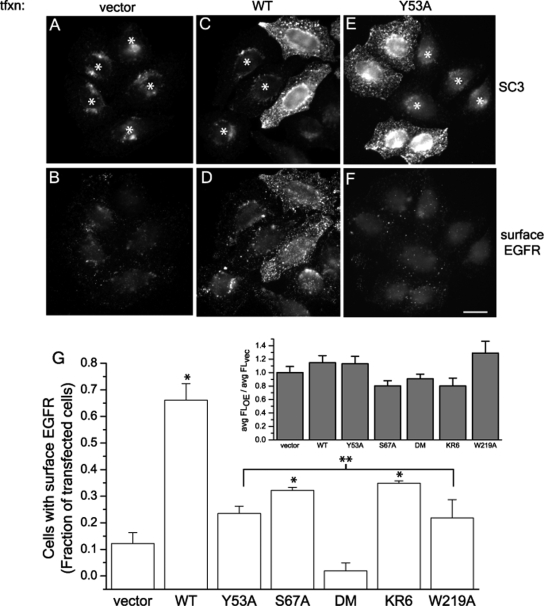
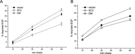
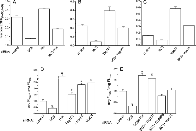
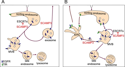
The epidermal growth factor receptor (EGFR) is targeted for lysosomal degradation by ubiquitin-mediated interactions with the ESCRTs (endosomal-sorting complexes required for transport) in multivesicular bodies (MVBs). We show that secretory carrier membrane protein, SCAMP3, localizes in part to early endosomes and negatively regulates EGFR degradation through processes that involve its ubiquitylation and interactions with ESCRTs. SCAMP3 is multimonoubiquitylated and is able to associate with Nedd4 HECT ubiquitin ligases and the ESCRT-I subunit Tsg101 via its PY and PSAP motifs, respectively. SCAMP3 also associates with the ESCRT-0 subunit Hrs. Depletion of SCAMP3 in HeLa cells by inhibitory RNA accelerated degradation of EGFR and EGF while inhibiting recycling. Conversely, overexpression enhanced EGFR recycling unless ubiquitylatable lysines, PY or PSAP motifs in SCAMP3 were mutated. Notably, dual depletions of SCAMP3 and ESCRT subunits suggest that SCAMP3 has a distinct function in parallel with the ESCRTs that regulates receptor degradation. This function may affect trafficking of receptors from prelysosomal compartments as SCAMP3 depletion appeared to sustain the incidence of EGFR-containing MVBs detected by immunoelectron microscopy. Together, our results suggest that SCAMP3, its modification with ubiquitin, and its interactions with ESCRTs coordinately regulate endosomal pathways and affect the efficiency of receptor down-regulation.


Figures

Figure 1.
Figure 1.
SCAMP3 contains conserved PY and P(S/T)AP motifs and is multimonoubiquitylated. (A) Organization of SCAMP3 featuring its proline-rich segment containing PY, PSAP, and PTEP motifs. (B) Interaction of SCAMP3 N-terminus with WW domain-containing proteins in a yeast two-hybrid screen; two Nedd4 ubiquitin ligases, Nedd4 and WWP2, and a third protein, YAP65, were identified as interaction partners. Mutation of SCAMP3's PY motif to PPAA (SC3Y53A) but not mutation of a distal PLPP (PAPP, SC3L61A) abrogated the interaction. (C) Pulldowns of SCAMP3 from lysates of rat parotid gland using GST fusions of mouse Nedd4 WW domains (WW1-3), corresponding mutant WW domains, and fusion protein made from Nedd4 cDNA retrieved in yeast two-hybrid screen (N4-Y2H). (D) Coimmunoprecipitation of untagged Nedd4 and FLAG-tagged Nedd4-2 expressed in HEK-293T cells using anti-SCAMP3 antibody or control IgG followed by immunoblotting with Nedd4 or FLAG antibody. (E) Ubiquitylation of SCAMP3. Lysates of HA-ubiquitin–transfected cells were immunoprecipitated with isoform-specific SCAMP1, 2, or 3 antibodies and immunoblotted with HA antibody (top), pan-SCAMP mAb (middle), or SCAMP3 mAb (bottom). Asterisk indicates ubiquitylated SCAMP3 species. (F) SCAMP3 is multimonoubiquitylated. Samples were immunoprecipitated as in E from lysates expressing wild-type or polymerization-defective HA-Ub KR4. The identical pattern of bands for Ub and UbKR4 demonstrates multimonoubiquitylation rather than addition of ubiquitin polymers of different length. Closed and open arrowheads denote the positions of SCAMP3 monomer and dimer, respectively, in C, E, and F.
Figure 2.
Figure 2.
Interaction of the PSAP motif in SCAMP3 with the UEV domain of Tsg101. (A) GST fusion proteins of SCAMP3 used in this study and calculated Kd from a representative binding experiment shown in B. n.d., not determined.
Figure 3.
Figure 3.
SCAMP3 interacts with Hrs. (A) HA-Hrs–transfected HEK-293T cells were lysed in the absence or presence of 10 mM N-ethylmaleimide (NEM), immunoprecipitated with anti-HA or control IgG, and immunoblotted with anti-SCAMP3 (top and middle panel) or anti-Hrs antibodies (bottom panel). The top panel is a short exposure, and the middle panel is a longer exposure of the same immunoblot to show ubiquitylated SCAMP3 species. (B). Mapping of the site on Hrs involved in SCAMP3 interaction. Lysates from cells expressing myc-tagged wild-type Hrs or indicated deletion mutants (ΔUIM, ΔVHS, ΔV/F, and ΔCBD) were immunoprecipitated with anti-myc antibody in the presence of 10 mM NEM and immunoblotted with anti-Hrs (top) or anti-SCAMP3 (bottom). (C) Interaction of the FYVE domain with SCAMP3. Lysates expressing GFP or GFP-2xFYVEHrs were immunoprecipitated with SCAMP3 antibody and immunoblotted with SCAMP3 mAb (top panels) or GFP (bottom panels). (D) Cells expressing myc-tagged Hrs were incubated for 30 min in the presence or absence of 100 ng/ml EGF, lysed, immunoprecipitated as in B, and immunoblotted with Hrs (top) or SCAMP3 antibody (bottom). (E) Endogenously expressed Hrs and SCAMP3 are partially colocalized. HeLa cells were permeabilized before fixation and immunostained for Hrs and SCAMP3. Bar, 10 μm.
Figure 4.
Figure 4.
Localization of SCAMP3 in relation to enlarged early endosomes. HeLa cells were transfected with GFP-Rab5Q79L (A) or GFP-2xFYVEHrs (B) or cotransfected with myc-Hrs and untagged mouse SCAMP3 (C). All samples were immunostained for SCAMP3; sample in C was also stained for myc. Enlargements of indicated areas are shown below the respective panels and illustrate focal localization of SCAMP3 along the membranes of expanded early endosomes stained by GFP (A and B) and substantial overlap with Hrs (C). Bar, 10 μm.
Figure 5.
Figure 5.
EGFR traffics to SCAMP3-rich compartments. HeLa cells were labeled with EGFR Ab 13A9, washed, and stimulated with 100 ng/ml EGF for 15, 30, or 60 min. Cells were then acid-stripped with low pH glycine to remove surface-associated EGFR antibody, fixed, permeabilized, and immunostained to detect internalized EGFR and SCAMP3. Bar, 10 μm.
Figure 6.
Figure 6.
RNAi-mediated knockdown of SCAMP3 enhances disappearance of fluorescent EGF. HeLa cells were transfected with scrambled, SCAMP1-, or SCAMP3-specific siRNA (no. 1), labeled with 100 ng/ml Alexa-488 EGF at 4°C, washed, and chased with 100 ng/ml EGF for indicated times at 37°C. (A) Representative images of cells transfected with scrambled or SCAMP3-specific siRNAs are shown; bar, 10 μm; asterisks at 60 min identify positions of cells in the images. (B) EGF fluorescence/cell was quantitated from Z-stacks of deconvolved images as described in Materials and Methods and was expressed as the average fluorescence ratio (knockdown/control) for each time point. The ratio for cells transfected with scrambled siRNA was set to 1.0. Error bars, SEM; * p < 0.01.
Figure 7.
Figure 7.
Knockdown of SCAMP3 accelerates EGF and EGFR degradation. Degradation (A) and recycling (B) of 125I-EGF in HeLa cells depleted of SCAMP3 (siRNA no. 2). Results from four independent experiments show that EGF degradation is accelerated and recycling inhibited compared with control cells. (C and D) Degradation of EGFR in HeLa cells transfected with control or SCAMP3-specific siRNA (no. 2). Cells were stimulated with 100 ng/ml EGF for indicated times, and the lysates were immunoblotted with specified antibodies. (C) A representative experiment is shown. (D) Quantitation of EGFR remaining after each time point from three independent experiments. EGFR levels were normalized for protein loading and quantitated as fraction of EGFR at time 0. Error bars, SEM.
Figure 8.
Figure 8.
Knockdown of SCAMP3 does not affect turnover and trafficking of transferrin receptor (TfR). (A) Surface-biotinylated cells were incubated for indicated times, lysed, adsorbed with streptavidin-agarose, and immunoblotted for TfR. The amount of TfR was normalized to the total biotinylated TfR (time 0). Results are averaged from three independent experiments and show no significant difference between control transfected or SCAMP3-depleted cells. Error bars, SEM. (B) Recycling of transferrin (125I-TfR) is the same in control transfected and SCAMP1- or 3-depleted HeLa cells. Recycling was measured in triplicate in three experiments as described in Materials and Methods. A representative experiment is shown. Error bars, SEM.
Figure 9.
Figure 9.
Recycling of EGFR is induced by SCAMP3 overexpression and is dependent on an intact PSAP motif and ubiquitylation. HeLa cells were transfected with vector (A and B), wild-type (WT) mouse SCAMP3 (C and D), SCAMP3 PY mutant Y53A (E and F), or other mutants not shown: PSAP mutant (S67A), double mutant (DM), ubiquitylation-deficient KR mutant (KR6), or W219A. Cells were surface labeled with EGFR Ab 13A9 and then stimulated with 100 ng/ml EGF for 2 h. After fixation, surface-associated EGFR was labeled with Alexa-594 secondary antibody (B, D, and F), and the cells were then permeabilized and immunostained for SCAMP3 using a low concentration of antibody to distinguish transfected from nontransfected cells (A, C, and E). Asterisk indicates nontransfected/low-expressing cells; bar, 10 μm. (G) Transfected cells were scored for the presence of surface-exposed EGFR relative to neighboring nontransfected cells. The average of 3–4 experiments is shown in the bar graph. * p < 0.05 compared with vector alone, ** p < 0.05 compared with wild-type (WT) transfected. Inset, internalization of EGF is not affected in overexpressing cells. Cells were labeled with 100 ng/ml Alexa-488 EGF at 4°C, washed, incubated with 100 ng/ml unlabeled EGF for 15 min at 37°C, and processed for immunostaining with a low concentration of SCAMP3 antibody. EGF fluorescence of SCAMP3 WT or mutant-expressing cells was quantitated as described in Materials and Methods, and results are expressed as average fluorescence ratio: SCAMP3 constructs overexpressed (OE):vector transfected (vec). Error bars, SEM.
Figure 10.
Figure 10.
Overexpression of wild-type SCAMP3 but not a double mutant inhibits EGF degradation and increases recycling. HeLa were transfected with vector (pcDNA 3.1), wild-type mouse SCAMP3, or DM-SCAMP3. (A) Degradation of 125I-EGF. (B) Recycling of 125I-EGF. Error bars, SEM, determined from three separate experiments.
Figure 11.
Figure 11.
Accelerated degradation of EGFR in SCAMP3-depleted cells is not blocked by simultaneous knockdown of ESCRT subunits. HeLa cells were transfected with control siRNA, SCAMP3 siRNA, and Hrs siRNA or Hrs and SCAMP3 siRNAs (A), Tsg101 siRNA or Tsg101 and SCAMP3 siRNAs (B), and Vps24 siRNA or Vps24 and SCAMP3 siRNAs (C). The cells were stimulated with 100 ng/ml EGF for 60 min, lysed, and immunoblotted for EGFR using ECL for quantitation. EGFR levels were normalized for protein levels and calculated as the fraction of total from time 0 (fraction EGFRt=60/t=0) in three independent experiments. Error bars, SEM. (D and E) EGF degradation in HeLa cells after knockdown of SCAMP3 and ESCRT subunits alone (D) and in paired combination (E). Cells were labeled with Alexa488-EGF and chased with 100 ng/ml EGF for 60 min. EGF fluorescence was quantitated as described in Materials and Methods, and results are expressed as average fluorescence ratio: knockdown:control transfected. Error bars, SEM; * p < 0.01.
Figure 12.
Figure 12.
EGFR-containing MVBs from SCAMP3-depleted cells have normal morphology. HeLa cells were transfected with control (A, B, E, F, and G) or SCAMP3 siRNA (C, D, H, I, and J), labeled with EGFR mAb 13A9 conjugated to gold, stimulated with 100 ng/ml EGF for 30 (A–D) or 60 min (E–J), and then processed for immunoelectron microscopy. Arrowheads indicate buds/tubules extending from the MVB. N, nucleus; LE, late endosome; bar, 0.25 μm.
Figure 13.
Figure 13.
Model of SCAMP3 function in inhibiting EGFR degradation and promoting recycling. After internalization, a fraction of EGFRs is sorted for lysosomal degradation in an ubiquitin and ESCRT-dependent manner. This sorting is negatively regulated by SCAMP3 through at least two possible mechanisms. (A) SCAMP3 inhibits ESCRT-mediated sorting of receptors into lysosomally directed MVBs and also promotes formation of a distinct population of MVBs destined for recycling. (B) Receptors are sorted into MVBs that mature en route to the lysosome. SCAMP3 inhibits ESCRT-mediated sorting of receptors into ILVs and facilitates their removal from maturing MVBs and trafficking through the endocytic recycling compartment (ERC).

References

    1. Alwan H. A., van Leeuwen J. E. UBPY-mediated epidermal growth factor receptor (EGFR) de-ubiquitination promotes EGFR degradation. J. Biol. Chem. 2007;282:1658–1669. - PubMed
    1. Amit I., et al. Tal, a Tsg101-specific E3 ubiquitin ligase, regulates receptor endocytosis and retrovirus budding. Genes Dev. 2004;18:1737–1752. - PMC - PubMed
    1. Babst M. A protein's final ESCRT. Traffic. 2005;6:2–9. - PubMed
    1. Bache K. G., Brech A., Mehlum A., Stenmark H. Hrs regulates multivesicular body formation via ESCRT recruitment to endosomes. J. Cell Biol. 2003a;162:435–442. - PMC - PubMed
    1. Bache K. G., Raiborg C., Mehlum A., Stenmark H. STAM and Hrs are subunits of a multivalent ubiquitin-binding complex on early endosomes. J. Biol. Chem. 2003b;278:12513–12521. - PubMed

Publication types

MeSH terms