. 2009 Mar;20(6):1749-62.
doi: 10.1091/mbc.e08-10-1033. Epub 2009 Jan 21.

Kinesin-5-dependent poleward flux and spindle length control in Drosophila embryo mitosis

Affiliations

Kinesin-5-dependent poleward flux and spindle length control in Drosophila embryo mitosis

Ingrid Brust-Mascher et al. Mol Biol Cell. 2009 Mar.

Abstract

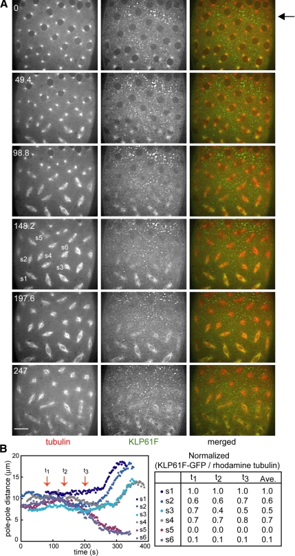
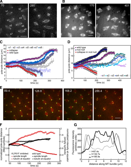
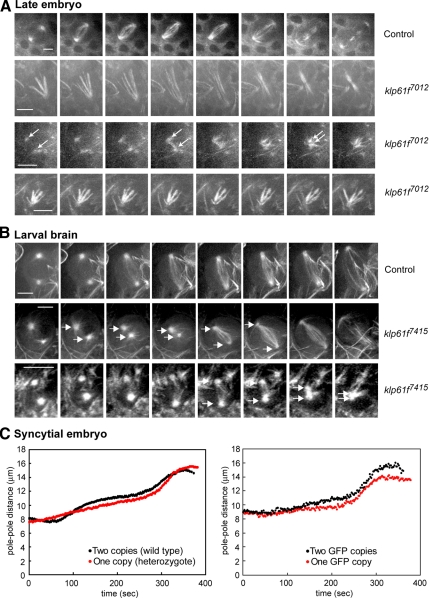
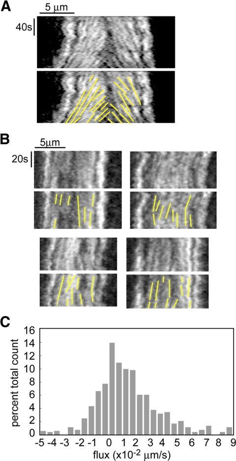
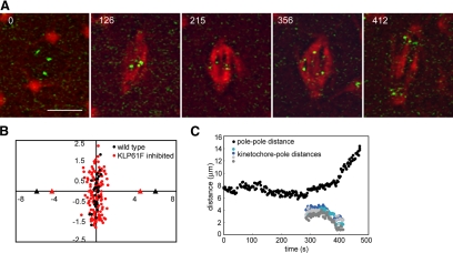
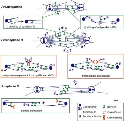
We used antibody microinjection and genetic manipulations to dissect the various roles of the homotetrameric kinesin-5, KLP61F, in astral, centrosome-controlled Drosophila embryo spindles and to test the hypothesis that it slides apart interpolar (ip) microtubules (MT), thereby controlling poleward flux and spindle length. In wild-type and Ncd null mutant embryos, anti-KLP61F dissociated the motor from spindles, producing a spatial gradient in the KLP61F content of different spindles, which was visible in KLP61F-GFP transgenic embryos. The resulting mitotic defects, supported by gene dosage experiments and time-lapse microscopy of living klp61f mutants, reveal that, after NEB, KLP61F drives persistent MT bundling and the outward sliding of antiparallel MTs, thereby contributing to several processes that all appear insensitive to cortical disruption. KLP61F activity contributes to the poleward flux of both ipMTs and kinetochore MTs and to the length of the metaphase spindle. KLP61F activity maintains the prometaphase spindle by antagonizing Ncd and another unknown force-generator and drives anaphase B, although the rate of spindle elongation is relatively insensitive to the motor's concentration. Finally, KLP61F activity contributes to normal chromosome congression, kinetochore spacing, and anaphase A rates. Thus, a KLP61F-driven sliding filament mechanism contributes to multiple aspects of mitosis in this system.


Figures

Figure 1.
Figure 1.
(A) Microinjection of an anti-KLP61F antibody (designed to dissociate KLP61F from spindles) results in a gradient of antibody concentration and produces a gradient in the KLP61F content of different spindles. Images from time-lapse movie of an embryo expressing KLP61F-GFP and injected with rhodamine tubulin and anti-KLP61F (see Supplemental Video 1). Time in each frame is given in seconds from NEB. Bar, 10 μm. The injection site was close to the top of the embryo (arrow), KLP61F-GFP forms immunoprecipitates, and most KLP61F-GFP is depleted from the spindles. These spindles collapse, as seen at 247 s. Toward the bottom of the embryo, KLP61F-GFP is still present on the spindles and consequently these spindles assemble, though they may exhibit defects. (B) Graph of pole–pole distance as a function of time (left) and quantification of KLP61F remaining on these spindles (right). The normalized ratio of KLP61F-GFP to rhodamine tubulin is used to compare the amount of motor remaining on each spindle at different time points. Spindles that collapse have practically no motor on them, whereas spindles that do not collapse or recover from partial collapse have at least 40% remaining.
Figure 2.
Figure 2.
(A) Two time points from a wild-type embryo injected with anti-KLP61F antibody showing a gradient of defects (see Supplemental Video 2). Bar, 10 μm. (B) Two time points from an Ncd null embryo injected with anti-KLP61F antibody showing more disorganized spindles due to the loss of two motors. Bar, 10 μm. (C) Graph of pole–pole distance as a function of time for the embryo shown in A. Close to the injection site spindles collapse (see s1 and s3), and other spindles shorten but remain small (see s2). Further away from the injection site, spindles do not elongate during prometaphase (s5, and the most distal spindles exhibit anaphase elongation (s6, s7, and s8). For comparison, we show graphs from a wild-type embryo (purple) and most severe effect (an embryo injected with enough antibody to collapse all spindles within the field of view; red). (D) Spindle length as a function of time for the Ncd null embryo shown in B. For comparison graphs of wild-type embryos (purple), Ncd null embryos (dark blue), and Ncd null embryos injected with a high concentration of anti-KLP61F (red) are shown. The gradient of concentration of antibody leads to a gradient of spindle pole lengths. A high inhibition of KLP61F leads to spindle collapse; lower concentrations lead to less severe defects. At a certain concentration the spindle maintains a steady length (light purple). (E) Anti-KLP61F antibody causes spindle collapse at prometaphase without apparent disruption of centrosomes or spindle poles. Images from time-lapse movie of an embryo expressing GFP-CNN (green) and injected with rhodamine tubulin (red) and anti-KLP61F (see Supplemental Video 3). Time in each frame is given in seconds. Bar, 10 μm. Note how the centrosomes move toward each other, but do not appear disorganized. (F) Tubulin fluorescence in the central spindle increases as the spindle collapses. Normalized spindle length (filled symbols) and fluorescent tubulin intensity at the equator (empty symbols) as a function of time for wild-type (black) and KLP61F inhibited spindles (red). After KLP61F inhibition spindles collapse with a correlated increase in tubulin intensity, suggesting that MTs are sliding inward and not depolymerizing. The increase is much higher than that observed in wild-type spindles, indicating that it is not due to a normal increase in tubulin. (G) Linescans of fluorescent tubulin intensity along a single ipMT bundle at different time points during the collapse. The intensity increases as the spindle collapses.
Figure 3.
Figure 3.
(A) Still images from real-time recordings of spindle pole separation dynamics in klp61f7012 mutant embryos expressing GFP-tubulin. Top row, spindle in wild-type embryo. Second row, a monoastral spindle that becomes bipolar. Third row, a spindle that collapses to form a permanent monoaster. Arrows mark the poles. Fourth row, monoaster that does not change during observation time. Bar, 5 μm. (B) Still images from real-time recordings of spindle pole separation dynamics in wild-type and klp61f7415 mutants expressing GFP-tubulin. Top row, a wild-type neuroblast. Second row, a mutant neuroblast with separated spindle poles that transiently collapse, then recover and separate fully. Third row, a mutant larval spindle that collapses to form a permanent monoaster. Bar, 5 μm. (C) Differences in spindle pole separation due to varying KLP61F gene dosage. Left, embryos from heterozygous mothers have a shorter metaphase length than wild-type embryos. Right, klp61f mutant embryos rescued with two copies of a KLP61F-GFP transgene have a longer metaphase spindle length than those rescued with one copy.
Figure 4.
Figure 4.
Poleward flux requires the function of KLP61F. (A) In wild-type embryos kymographs show that tubulin speckles flux toward the poles during the metaphase–anaphase A steady state, but move at the same rate as the poles during anaphase B. (Brust-Mascher and Scholey, 2002). (B) In embryos injected with anti-KLP61F antibody, kymographs obtained from spindles that did not collapse but maintained a steady length show that tubulin speckles do not flux toward the pole, indicating that the function of KLP61F is required for this movement (see Supplemental Video 2 for speckled movie). (C) Histogram of all the flux data obtained on spindles that did not collapse after KLP61F inhibition.
Figure 5.
Figure 5.
Chromosome congression and anaphase A are perturbed by partial KLP61F inhibition. (A) Time-lapse images of one spindle in an embryo expressing GFP-CID injected with rhodamine tubulin and anti-KLP61F antibody. The spindle shortens slightly (compare 215 with 126) but does not collapse after KLP61F inhibition. Kinetochores move to the poles albeit slower than in control spindles. Time in each frame is given in seconds from NEB. Bar, 5 μm. (B) Positions of the center of kinetochore pairs in wild-type (black) and KLP61F-inhibited (red) spindles. In anti-KLP61F–inhibited spindles, kinetochores do not congress as tightly as in controls, and they exhibit a larger displacement around the equator. Triangles mark the average positions of the poles. (C) Pole–pole distance and kinetochore-to-pole distances for the spindle shown in A (averages in Table 1).
Figure 6.
Figure 6.
Model for the multiple roles of KLP61F in Drosophila syncytial embryo mitosis. We propose that kinesin-5 motors function as ensembles of dynamic, transient, MT crosslinkers throughout the spindle (Sharp et al., 1999a; Cheerambathur et al., 2008), organizing parallel MTs into bundles (inset i) and sliding apart antiparallel MTs (inset ii), in accordance with the biochemical properties of KLP61F (e.g., Cole et al., 1994; Kashina et al., 1996a; Tao et al., 2006; Van den Wildenberg et al., 2008). The results suggest that KLP61F-driven MT-MT sliding is required to oppose an inward force (ii, brown arrows) and maintain the spindle during early prometaphase, as well as to elongate it to the metaphase steady state length. In preanaphase B (i.e. metaphase and anaphase A) spindles, KLP61F driven ipMT outward sliding (blue solid arrows) is required for poleward flux in both kMT and ipMT bundles and for anaphase A chromosome movement. The kMTs may be slid poleward (dashed blue arrows) via crosslinks to the actively sliding antiparallel ipMTs. Finally, KLP61F drives spindle elongation during anaphase B. This model is proposed only for Drosophila embryos (see Discussion).

References

    1. Bettencourt-Dias M., et al. Genome-wide survey of protein kinases required for cell cycle progression. Nature. 2004;432:980–987. - PubMed
    1. Blower M. D., Karpen G. H. The role of Drosophila CID in kinetochore formation, cell-cycle progression and heterochromatin interactions. Nat. Cell Biol. 2001;3:730–739. - PMC - PubMed
    1. Brust-Mascher I., Civelekoglu-Scholey G., Kwon M., Mogilner A., Scholey J. M. Model for anaphase B: role of three mitotic motors in a switch from poleward flux to spindle elongation. Proc. Natl. Acad. Sci. USA. 2004;101:15938–15943. - PMC - PubMed
    1. Brust-Mascher I., Scholey J. M. Microtubule flux and sliding in mitotic spindles of Drosophila embryos. Mol. Biol. Cell. 2002;13:3967–3975. - PMC - PubMed
    1. Brust-Mascher I., Scholey J. M. Mitotic spindle dynamics in Drosophila. Int. Rev. Cytol. 2007;259:139–172. - PubMed

Publication types

LinkOut - more resources