. 2009 Mar;20(6):1865-77.
doi: 10.1091/mbc.e08-08-0838. Epub 2009 Jan 21.

The two SAS-6 homologs in Tetrahymena thermophila have distinct functions in basal body assembly

Affiliations

The two SAS-6 homologs in Tetrahymena thermophila have distinct functions in basal body assembly

Brady P Culver et al. Mol Biol Cell. 2009 Mar.

Abstract

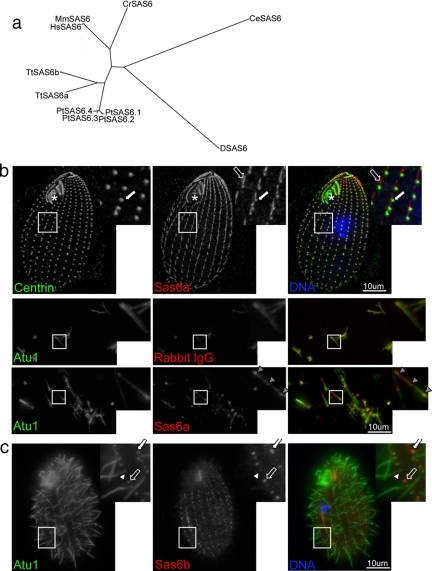
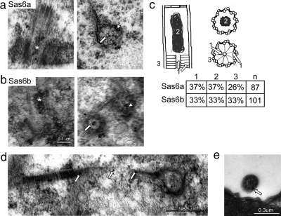
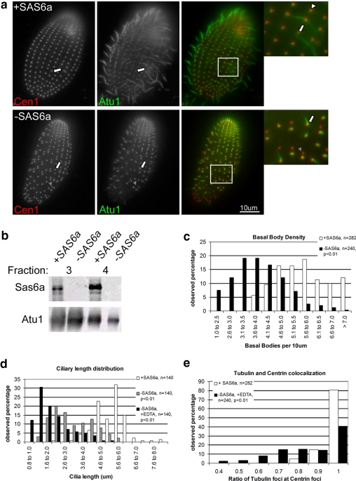
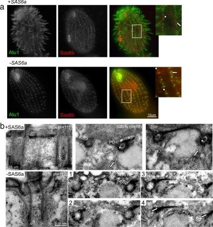
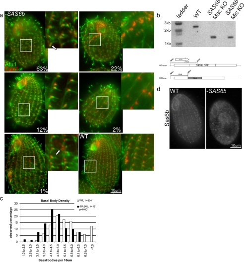
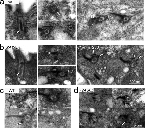
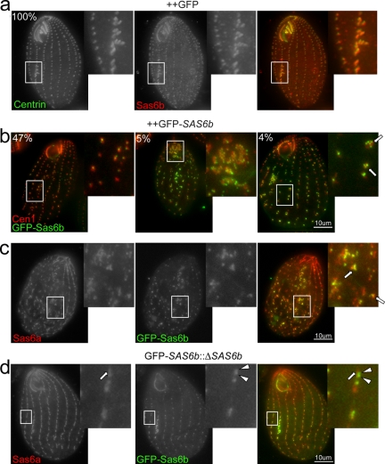
Cilia and flagella are structurally and functionally conserved organelles present in basal as well as higher eukaryotes. The assembly of cilia requires a microtubule based scaffold called a basal body. The ninefold symmetry characteristic of basal bodies and the structurally similar centriole is organized around a hub and spoke structure termed the cartwheel. To date, SAS-6 is one of the two clearly conserved components of the cartwheel. In some organisms, overexpression of SAS-6 causes the formation of supernumerary centrioles. We questioned whether the centriole assembly initiation capacity of SAS-6 is separate from or directly related to its structural role at the cartwheel. To address this question we used Tetrahymena thermophila, which expresses two SAS-6 homologues, TtSAS6a and TtSAS6b. Cells lacking either TtSAS6a or TtSAS6b are defective in new basal body assembly. TtSas6a localizes to all basal bodies equally, whereas TtSas6b is enriched at unciliated and assembling basal bodies. Interestingly, overexpression of TtSAS6b but not TtSAS6a, led to the assembly of clusters of new basal bodies in abnormal locations. Our data suggest a model where TtSAS6a and TtSAS6b have diverged such that TtSAS6a acts as a structural component of basal bodies, whereas TtSAS6b influences the location of new basal body assembly.


Figures

Figure 1.
Figure 1.
Conservation and localization of Tetrahymena Sas6 proteins. (a) Dendrogram showing relative distances between SAS-6 genes based on PISA domain alignment. Tt, Tetrahymena thermophila TTHERM_00388200 (TtSAS6a) and TTHERM_00137600 (TtSAS6b); Pt, Paramecium tetraurelia GSPATP00011824001 (PtSAS6.1); GSPATP00008149001 (PtSAS6.2), GSPATP00005603001 (PtSAS6.3), and GSPATP00024268001 (PtSAS6.4); Hs, Homo sapiens GI:35038001; Mm, Mus musculus GI:62511043; Cr. Chlamydomonas reinhardtii GI:161727419; Ce. C. elegans GI:1754348; D, Drosophila melanogaster GI:62511095. (b) Sas6a localizes to basal bodies and kinetodesmal fibers in whole cells and to foci in isolated cilia preparations. Cells are oriented in this and all fluorescent images with anterior side up. Fixed wild-type Tetrahymena cells were labeled with antibodies to centrin (left panel, green in merge) and Sas6a (middle panel, red in merge) as well as DAPI stain to mark DNA (blue in merge). The asterisk indicates the oral apparatus, the white arrow shows centrin and Sas6a basal body localization, and the unfilled arrow shows Sas6a kinetodesmal fiber localization. In cilia fluorescent images, fixed isolated cilia preparations were labeled with antibodies to α-tubulin (Atu1, left panel, green in merge) and rabbit IgG or Sas6a (middle panels, red in merge). The gray arrowheads show Sas6a foci present along the length of the cilia. (c) Sas6b is enriched at unciliated and assembling basal bodies. Fixed wild-type Tetrahymena cells were labeled with antibodies to Atu1 (left panel, green in merge), Sas6b (middle panel, red in merge) as well as DAPI stain to mark nuclei. The white arrow shows an example of an assembling basal body, the unfilled arrow shows an unciliated basal body, and the white arrowhead shows a ciliated basal body.
Figure 2.
Figure 2.
Sas6a and Sas6b localize to all of the same structural domains within the basal body. (a) Sas6a localizes to the cartwheel hub, spokes, electron-dense lumen, and periphery of the basal body at the proximal end. Immunoelectron micrographs showing examples of Sas6a localization observed in whole cell preparations. The Sas6a antibody was visualized using a 25-nm gold–conjugated secondary antibody. The white asterisk indicates the electron-dense lumen, the arrow points to the hub of the cartwheel, the arrowhead shows cartwheel spoke to “A” tubule attachment point, and the black asterisk shows the basal body periphery associated Sas6a. (b) Sas6b shares all of the same localizations as Sas6a. The Sas6b antibody was visualized using a 25-nm gold–conjugated secondary antibody. Immunoelectron micrographs of whole cell preparations showing examples of Sas6b localizations, the basal body structural domains are indicated as in panel a. (c) Sas6a and Sas6b localize at nearly the same frequency to the same structural domains within the basal body. The schematic shows a basal body in longitudinal section (left drawing) as well as a view of the electron-dense lumen in cross section (right, top drawing) and the cartwheel (right, bottom drawing). Structural domains were counted as indicated in the drawings (1, cartwheel hub and spokes; 2, electron-dense lumen; 3, periphery of the basal body). The chart shows the frequency (n) gold particles were observed for each antibody at each position. (d) Sas6a localizes to the kinetodesmal fiber. The white arrows indicate anti-Sas6a particles found along the kinetodesmal fiber associated with a basal body in cross section (right). (e) Sas6a localizes to the periphery of cilia. The arrow indicates the position of a 25-nm gold particle.
Figure 3.
Figure 3.
SAS6a is required for new basal body assembly. (a) SAS6a is required for microtubule incorporation into basal bodies and for maintenance of cilia length. Fixed whole cells either grown in the presence (+SAS6a) or absence (−SAS6a) of SAS6a were labeled with antibodies to Cen1 (left panels, red in merge) and Atu1 (middle panels, green in merge). The white arrows show examples of a ciliated basal body, the white arrowhead shows an unciliated basal body, and the gray arrowhead shows a putative incomplete basal body: Cen1 signal is present, but Atu1 signal is absent. (b) SAS6a expression is effectively repressed by inclusion of 100 μM EDTA in the media of the SAS6a conditional null strain. Western blot showing levels of Sas6a and Atu1 in the indicated fractions for the indicated conditions. Equal total protein was loaded in this blot. Fraction 3, protein soluble in HpHS buffer (see Materials and Methods), and fraction 4, the insoluble pellet from this treatment. Sas6a was not found in fractions 1 and 2 in either condition (data not shown). (c) Graphical representation of the basal body assembly defect seen upon activation or repression of SAS6a. Basal bodies were counted based on instances of Cen1 and Atu1 colocalization. Graph shows the frequency of basal body density observed in a population of cells grown with (+SAS6a) or without (−SAS6a) SAS6a. −SAS6a cells have significantly fewer basal bodies per 10 μm than +SAS6a cells. (d) Cells grown without induction of SAS6a display shorter cilia than cells grown with induction of SAS6a. Graphical representation of the distribution of cilium length seen in a population of cells grown with active repression of SAS6a (−SAS6a, +EDTA), incomplete repression of SAS6a (−SAS6a), or with expression of SAS6a (+SAS6a). Cilia were measured from the base to the tip and plotted as the frequency of range of lengths observed in the population. (e) Loss of SAS6a results in a significant increase of aberrant basal bodies. Graphical representation of the frequency at which Cen1 foci were observed with Atu1 foci shows that cells grown in the absence of SAS6a (−SAS6a) had fewer instances of Cen1 and Atu1 colocalization than cells grown with SAS6a (+SAS6a). The number of Cen1 foci colocalizing with Atu1 foci were counted along individual ciliary rows for these calculations.
Figure 4.
Figure 4.
SAS6a is required for the maintenance of the cartwheel structure. (a) Fixed cells grown in the presence (+SAS6a) or absence (−SAS6a) were labeled with antibodies to Atu1 (left panels, green in merge) and Sas6b (middle panels, red in merge). Sas6b signal at unciliated or assembling basal bodies (arrowheads) was on average 2.9-fold greater than Sas6b signal at ciliated basal bodies (arrows) when SAS6a was expressed (n = 149). When SAS6a expression was repressed (−SAS6a), this ratio decreased to 1.2 (n = 151). (b) Repression of SAS6a is correlated with the loss of the cartwheel from mature basal bodies and the appearance of ribosomes in lumen of the basal body. Electron micrographs of whole cell preparations showing basal bodies in longitudinal section (left panels) or cross section (right panels) grown with (+SAS6a) or without (−SAS6a). +SAS6a basal bodies have the cartwheel (unfilled arrows), the electron-dense lumen (white arrowhead), and the markers of mature basal bodies: the postciliary microtubules (PC) and kinetodesmal fibers (KF). Ribosomes (white arrows) are excluded from the basal body lumen. −SAS6a basal bodies often lacked a complete cartwheel (unfilled arrows), and loss of the cartwheel was correlated with the appearance of ribosomes in the lumen of the basal body. The percentages indicate the frequency at which basal bodies like the ones in the figure were observed (see Materials and Methods). Examination of serial sections of two basal bodies in cross section (1, most proximal; 4, most distal) demonstrates this. Structural features are indicated as in +SAS6a condition.
Figure 5.
Figure 5.
SAS6b is important for but not essential to vegetative cell growth and new basal body assembly. (a) Loss of SAS6b is correlated with appearance of cells with fewer basal bodies and/or lacking complete oral apparatus. Fixed wild-type (WT) and SAS6b KO cells (−SAS6b) were labeled with antibodies to Cen1 (left panels, red merge) and Atu1 (middle panels, green merge). Cells (n = 300) that resembled the cells in the images shown in this figure were counted. The percentage of cells that matched these representative images is shown in the bottom right-hand corner. The arrow in the first cell (63%) indicates a branching of a cortical row. The arrow in the fifth panel (1%) indicates a cluster of basal bodies present along a cortical row. (b) SAS6b is not required for vegetative cell growth. Southern blot showing genomic DNA from indicated strains digested with XhoI and NheI. The blot was probed with DNA 1 kb 5′ of the SAS6b start site. Diagram below blot shows wild-type (WT) SAS6b locus and null (KO) locus. The wild-type fragment recognized by the probe is 2.3 kb and the null fragment is 1.3 kb. (c) SAS6b null cells have fewer basal bodies per 10 μm than wild type. Basal bodies were counted based on instances of Cen1 and Atu1 colocalization. (d) Sas6b basal body signal is absent in SAS6b KO cells. Fixed wild-type (WT) or SAS6b KO cells (−SAS6b) were labeled with the anti-Sas6b antibody.
Figure 6.
Figure 6.
Loss of SAS6b does not affect cartwheel assembly, but does result in accumulation of electron densities near the site of new basal body assembly. (a) Electron micrographs showing wild-type (WT) basal bodies in longitudinal or cross section. Arrowhead, the electron-dense lumen; unfilled arrow, a cartwheel in cross section. A new basal body (asterisk) is seen in the panel on the right just below the kinetodesmal fiber (KF) and opposite of the postciliary microtubules (PC). (b) Basal bodies from SAS6b null cells (−SAS6b) possess all of the structures associated with wild-type basal bodies. Relevant structures are indicated as in panel a. Fifty-one percent of basal bodies examined (n = 200) showed electron densities (arrows) at or near the site of new assembly. (c) Cen1 localizes to the site of new basal body assembly in wild-type (WT) cells. Immunoelectron micrographs using a 25-nm gold–conjugated secondary antibody showing the localization of Cen1. The arrow indicates Cen1 localization at the site of new assembly. (d) Cen1 localizes to the site of new assembly in SAS6b null cells (−SAS6b) and is associated with the electron densities. Immunoelectron micrographs labeled as in panel c. The arrows point to Cen1 association with the electron densities.
Figure 7.
Figure 7.
SAS6b helps dictate sites of new basal body assembly and acts upstream of SAS6a. (a) Overexpression of GFP does not affect cellular morphology. Fixed cells overexpressing GFP (++GFP) were labeled with antibodies to centrin (left panel, green in merge) and Sas6b (middle panel, red in merge). Sas6b is enriched at the developing oral apparatus (inset), which is always found immediately below the existing oral apparatus along the equator of the cell (100%, n = 200). (b) Overexpression of GFP-SAS6b causes the formation of clusters of basal bodies in abnormal locations. Fixed cells overexpressing GFP-SAS6b were labeled with antibodies to Cen1 (red) and GFP (green). Forty-seven percent of cells in the population had disorganized cortical rows and large gaps between existing basal bodies (left panel). Five percent of cells had an abnormal oral apparatus (middle panel), and 4% of cells had clusters of basal bodies in abnormal locations (right panel, n = 203). Arrows point to clusters of basal bodies in abnormal locations. (c) Sas6a colocalizes with GFP-Sas6b clusters. Fixed cells overexpressing GFP-SAS6b were labeled with antibodies to Sas6a and GFP. Arrows indicate Sas6a and GFP colocalization. (d) GFP-Sas6b localizes to presumptive sites of new basal body assembly before Sas6a. Fixed SAS6b macronuclear null cells carrying an inducible GFP-SAS6b allele at a different locus were induced to express GFP-SAS6b and labeled with antibodies to Sas6a (left panel, red in merge) and GFP (middle panel, green in merge). The arrow shows Sas6a kinetodesmal fiber localization. The arrowheads show GFP-Sas6b doublets representing a new basal body assembly event.

References

    1. Allen R. D. The morphogenesis of basal bodies and accessory structures of the cortex of the ciliated protozoan Tetrahymena pyriformis. J. Cell Biol. 1969;40:716–733. - PMC - PubMed
    1. Badano J. L., Mitsuma N., Beales P. L., Katsanis N. The ciliopathies: an emerging class of human genetic disorders. Annu. Rev. Genomics Hum. Genet. 2006;7:125–148. - PubMed
    1. Brown T. Southern blotting. In: Ausubel F. M., Brent R., Kingston R. E., Moore D. D., Seidman J. G., Smith J. A., Struhl K., editors. Current Protocols in Molecular Biology. Vol. 1. Hoboken, NJ: John Wiley and Sons; 1999. pp. 2.9.1–2.9.15.
    1. Bruns P. J., Cassidy-Hanley D. Biolistic transformation of macro- and micronuclei. Methods Cell Biol. 2000;62:501–512. - PubMed
    1. Callaini G., Whitfield W. G., Riparbelli M. G. Centriole and centrosome dynamics during the embryonic cell cycles that follow the formation of the cellular blastoderm in Drosophila. Exp. Cell Res. 1997;234:183–190. - PubMed

Publication types

Substances

LinkOut - more resources