. 2009 Mar;20(6):1705-14.
doi: 10.1091/mbc.e08-03-0282. Epub 2009 Jan 21.

The SM protein Car/Vps33A regulates SNARE-mediated trafficking to lysosomes and lysosome-related organelles

Affiliations

The SM protein Car/Vps33A regulates SNARE-mediated trafficking to lysosomes and lysosome-related organelles

Mohammed A Akbar et al. Mol Biol Cell. 2009 Mar.

Abstract

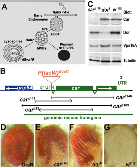
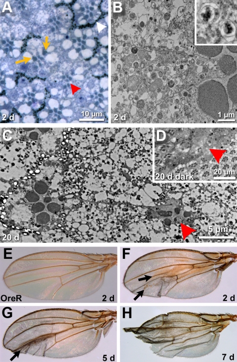
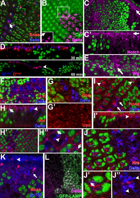
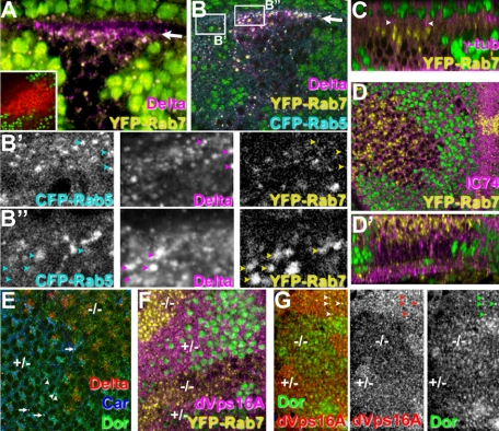
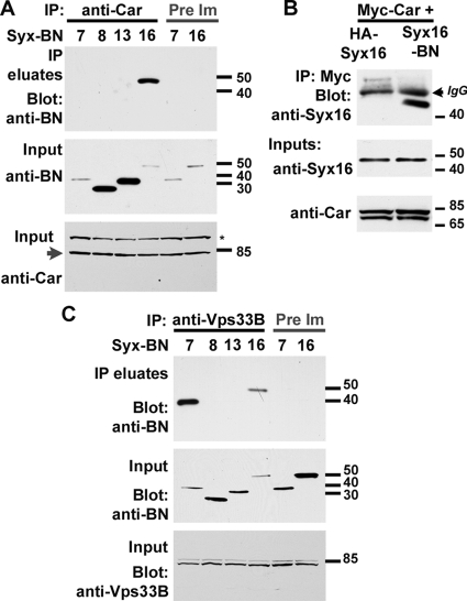
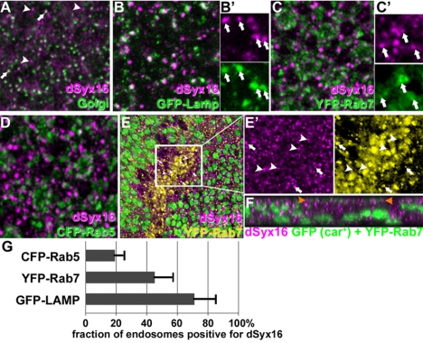
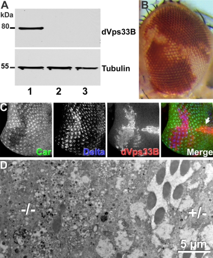
The SM proteins Vps33A and Vps33B are believed to act in membrane fusions in endosomal pathways, but their specific roles are controversial. In Drosophila, Vps33A is the product of the carnation (car) gene. We generated a null allele of car to test its requirement for trafficking to different organelles. Complete loss of car function is lethal during larval development. Eye-specific loss of Car causes late, light-independent degeneration of photoreceptor cells. Earlier in these cells, two distinct phenotypes were detected. In young adults, autophagosomes amassed indicating that their fusion with lysosomes requires Car. In eye discs, endocytosed receptors and ligands accumulate in Rab7-positive prelysosomal compartments. The requirement of Car for late endosome-to-lysosome fusion in imaginal discs is specific as early endosomes are unaffected. Furthermore, lysosomal delivery is not restored by expression of dVps33B. This specificity reflects the distinct pattern of binding to different Syntaxins in vitro: dVps33B predominantly binds the early endosomal Avl and Car to dSyntaxin16. Consistent with a role in Car-mediated fusion, dSyntaxin16 is not restricted to Golgi membranes but also present on lysosomes.


Figures

Figure 1.
Figure 1.
Car is an essential gene. (A) Cartoon shows endocytic intermediates and the markers used in this study for their detection. (B) Diagram of the car genomic region. Imprecise excision of the P-element P[lacW]G0447 in the 5′ UTR of the car yielded the indicated deletions. Molecular analysis revealed that carΔ146 removed almost the entire car coding unit without extending into the grip84 gene. The carΔ146 deletion was used for further studies. Lethality of the carΔ146 chromosome was rescued by the indicated genomic rescue construct. (C) Western blot of lysates (20 μg of protein) from carΔ146, dor8 and w1118 second instars were probed with antibodies against Car, Dor, dVps16A, or α-tubulin as control. (D–G) Micrographs of adult eyes reveal reduced pigmentation of clones of carΔ146 (E) or dor8 (F) homozygous mutant cells compared with Oregon R (D) or w1118 (G) control eyes. Genotypes: carΔ146, dor8, and w1118 (C), Oregon R (D), w+ carΔ146 FRT19A/w+ FRT19A; hsFLP (E), w+ dor8 FRT19A/w+ FRT19A; hsFLP (F), w1118 (G).
Figure 2.
Figure 2.
Loss of car causes late degeneration. (A) Micrograph of a section of a 2-d-old adult eye containing carΔ146 mutant cells identified by the loss of pigmentation. The trapezoidal arrangement of rhabdomeres characteristic for wild-type ommatidia (white arrowhead) is not altered in the carΔ146ommatidia (red arrowhead). Rhabdomeres of mutant cells are smaller. This phenotype is cell autonomous as in mixed ommatidia car+ photoreceptor cells (recognized by their normal pigmentation, orange arrows) also have normal rhabdomere size but do not rescue neighboring carΔ146 cells (identified by their lack of pigmentation in a w+ background. (B) An electron micrograph of a 2-d-old carΔ146 ommatidium reveals the accumulation of vacuoles in the mutant photoreceptor cells, which nevertheless display well-organized rhabdomeres. The inset shows two examples in which the double membranes typical for autophagosomes are resolved. (C) Electron micrograph of a 20-d-old compound eye. A carΔ146 ommatidium (red arrowhead) shows massive degeneration. (D) A micrograph depicts the eye of a fly raised for 20 d in the dark. Mutant ommatidia are degenerated (red arrowhead). (E–H) Wings from wild-type flies (E) or flies carrying carΔ146 clones. Wing clones are not marked, but the majority of flies exhibiting carΔ146 clones in the eye contained brown blemishes on the wings after just 2 to 5 d (arrows in F and G). Later, most of those wings became frail and damaged (H). Genotypes: w+ carΔ146 FRT19A/w+ FRT19A; hsFLP (A–D and F–H) Oregon R (E).
Figure 3.
Figure 3.
Car is required for normal trafficking of endocytosed proteins. In micrographs of parallel (A–C, E–K) or Z-sections (C′, D, H, and I′) the absence of the nuclear GFP marker (A, C–K) or Car staining (B) identified clones of carΔ146 mutant cells in otherwise wild-type eye discs or wing discs (E). In carΔ146 cells, Boss and Delta (A), Delta (B), Notch (C), and Wg (E) accumulated. The inset in B shows the Delta channel only to visualize the comparably low level of Delta in wild-type cells (arrow). Levels of apical cell surface Notch protein (arrows in C and C′) were not altered despite the intracellular accumulation. (D) Antibodies against the extracellular domain of Delta (Dextra) were internalized by live eye discs for the indicated time. Subsequently, discs were fixed, permeabilized and the location of internalized antibodies was compared with that of total Delta (Dtotal). The arrowhead points to examples of antibodies internalized into the large Delta-positive compartments in carΔ146 clones. (F) Delta-positive compartments in carΔ146 clones were negative for the Golgi marker Lvl. (G) Upd was not up-regulated in carΔ146 clones. Large Delta-positive compartments in carΔ146 clones (arrowheads in H–J) were negative for the endosomal markers Avl (H), Hook (I), and Hrs (J), but in wild-type and carΔ146 mutant cells small apical Delta-positive vesicles were positive for these endosomal markers (arrows in H, H”, and J′). (K) The late endosomal protein gp150 colocalizes with Boss proteins endocytosed into carΔ146 mutant R7 cells (arrow). (L) Delta-positive compartments were labeled by GFP-LAMP1, which accumulated in carΔ146 cells. Genotypes: w1118 carΔ146 FRT19A/w1118 P[w+,ubi>nGFP] FRT19A; hsFLP (A–K).
Figure 4.
Figure 4.
Loss of car function causes the accumulation of cargo in displaced Rab7-positive late endosomes. The absence of a nuclear GFP marker (green in A–D, F, and G) or Car staining (E) identified carΔ146 cells in otherwise wild-type wing discs (A, B, F, and G) or eye discs (C–E). (A and B) Z-sections of imaginal discs taken at a slight angle revealed that in carΔ146 mutant cells, Delta accumulated in YFP-Rab7–positive late endosomes, which were highly enriched just below the apical surface (arrows in A and B). The inset in A shows the accumulation of LysoTracker (red) in acidified endosomes of carΔ146 mutant cells (marked by absence the nuclear GFP). Higher magnification of CFP-Rab5, Delta and YFP-Rab7 depict Delta in Rab5-positive early endosomes in wild-type cells (B′) but accumulated in Rab7-positive late endosomes in carΔ146 mutant cells (B″). (C) YFP-Rab7–positive late endosomes are enriched close to centrosomes marked by γ-tubulin and are labeled by IC74 (D), which recognizes the dynein IC (D′ is a Z-section). (E) In wild-type cells (+/−), Car and Dor colocalize (examples labeled by arrowheads) and are found on some vesicles containing Delta (arrows). However, in carΔ146 cells Dor is not reduced but does not label the large Delta-positive vesicles. (F) In carΔ146 cells (−/−) with apical enriched YFP-Rab7 endosomes, apical dVps16A is reduced. (G) Dor and dVps16A colocalize in wild-type cells (arrowheads), but in carΔ146 cells (−/−) dVps16A is reduced, whereas Dor seems enhanced. Genotypes: w1118 carΔ146 FRT19A/w1118 P[w+,ubi>nGFP] FRT19A; hsFLP; P[w+ tub>YFP-Rab7] (A, C, D, and F); w1118 carΔ146 FRT19A/w1118 P[w+,ubi>nGFP] FRT19A; hsFLP; P[w+ tub>YFP-Rab7] P[w+ tub>CFP-Rab5] (B) w1118 carΔ146 FRT19A/w1118 FRT19A; hsFLP (E) w1118 carΔ146 FRT19A/w1118 P[w+,ubi>nGFP] FRT19A; hsFLP (G).
Figure 5.
Figure 5.
Car binds to dSyntaxin16. Car (A and B) or dVps33B (C) were coexpressed in S2 cells with the indicated Syntaxins carrying a C-terminal BN epitope-tag replacing the transmembrane domain or an N-terminal HA-tag in (B). Expression levels were detected on blots of input samples by using the indicated antibodies. Protein complexes were immunoprecipitated with anti-Car (A), anti-Myc (B), or anti-dVps33B (C) antibodies or preimmunsera when indicated. Coimmunoprecipitated syntaxins were detected using the anti-BN antibodies (A and C) or anti-dSyntaxin16 (B). Note that dSyntaxin7 is synonymous with Avl.
Figure 6.
Figure 6.
dSyntaxin16 is present in Golgi and lysosomes. Micrographs of eye discs visualize dSyntaxin16 and markers for the Golgi complex (A; 7H6D7C2), lysosomes (B; GFP-Lamp), late endosomes (C; YFP-Rab7), and early endosomes (D; CFP-Rab5). Although dSyntaxin16 extensively colocalized with the Golgi marker (arrows in A) a subset of dSyntaxin16-positive punctae is negative for 7H6D7C2 staining (arrowheads in A). Some dSyntaxin16-positive punctae colocalized with GFP-Lamp (arrows in B′) or YFP-Rab7 (arrows in C′) but rarely CFP-Rab5 (D). In carΔ146 mutant cells, YFP-Rab7-positive late endosomes were enriched but negative for dSyntaxin16 (E and F). Higher magnifications micrographs depict the colocalization of YFP-Rab7 and dSyntxin16 in neighboring wild-type cells (arrows in E′) but in carΔ146 cells the few strongly stained dSyntaxin16 punctae were negative for YFP-Rab7 (arrowheads in E′). The Z-section depicted in F shows a carΔ146 clone. Between the arrowheads apically accumulated YFP-Rab7 is shown in green. (G) Quantification of the fraction of Rab5-marked early endosomes, Rab7-marked late endosomes or Lamp-marked lysosomes that were also stained for endogenous dSyx16. Genotypes: OreR (A) w1118; [w+,tub>GFP-LAMP] (B) w1118; [w+,tub>YFP-Rab7] (C) w1118; [w+,tub>CFP-Rab5] (D) w1118 carΔ146 FRT19A/w1118 P[w+,ubi>nGFP] FRT19A; hsFLP; P[w+ tub>YFP-Rab7] (E).
Figure 7.
Figure 7.
dVps33B cannot replace Car. (A) Western blots of lysates from fly heads with the genotypes (1) carΔ146 FRT19A/tub-Gal80 FRT19A; tub-Gal4; UAS-Myc-dVps33B (2) UAS-Myc-dVps33B (3) carΔ146 FRT19A/tub-Gal80 FRT19A; tub-Gal4 show the expression of Myc-dVps33B in MARCM clones (Lee and Luo, 2001). (B) Micrograph of an adult eye expressing dVps33B by using the MARCM technique, which did not restore pigmentation in carΔ146 mutant cells. (C) Expression of dVps33B in a carΔ146 clone did not restore normal trafficking of Delta in a third instar eye disk. (D) An electron micrograph of a section of an adult eye shows that dVps33B expression did not prevent degeneration of carΔ146 cells. Genotypes: w+ carΔ146 FRT19A/w1118 P[w+ tubP>GAL80] hsFLP, FRT19A; P[w+ tubP>Gal4]/+; P[w+ UAS-Myc-dVps33B]/+ (B–D).

References

    1. Aikawa Y., Lynch K. L., Boswell K. L., Martin T. F. A second SNARE role for exocytic SNAP25 in endosome fusion. Mol. Biol. Cell. 2006;17:2113–2124. - PMC - PubMed
    1. Banta L. M., Vida T. A., Herman P. K., Emr S. D. Characterization of yeast Vps33p, a protein required for vacuolar protein sorting and vacuole biogenesis. Mol. Cell Biol. 1990;10:4638–4649. - PMC - PubMed
    1. Beadle G. W., Ephrussi B. The differentiation of eye pigments in Drosophila as studied by transplantation. Genetics. 1936;21:225–247. - PMC - PubMed
    1. Bethani I., Lang T., Geumann U., Sieber J. J., Jahn R., Rizzoli S. O. The specificity of SNARE pairing in biological membranes is mediated by both proof-reading and spatial segregation. EMBO J. 2007;26:3981–3992. - PMC - PubMed
    1. Boehm M., Bonifacino J. S. Adaptins: the final recount. Mol. Biol. Cell. 2001;12:2907–2920. - PMC - PubMed

Publication types

MeSH terms

LinkOut - more resources