. 2009 Jan 1;8(1):158-66.
doi: 10.4161/cc.8.1.7533.

Molecular phenotyping of human ovarian cancer stem cells unravels the mechanisms for repair and chemoresistance

Affiliations

Molecular phenotyping of human ovarian cancer stem cells unravels the mechanisms for repair and chemoresistance

Ayesha B Alvero et al. Cell Cycle. .

Abstract

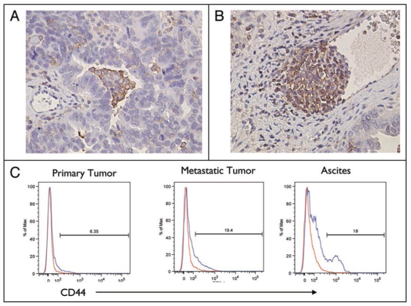
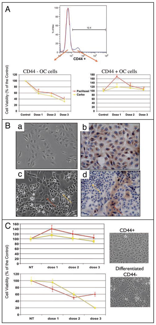
A major burden in the treatment of ovarian cancer is the high percentage of recurrence and chemoresistance. Cancer stem cells (CSCs) provide a reservoir of cells that can self-renew, can maintain the tumor by generating differentiated cells [non-stem cells (non-CSCs)] which make up the bulk of the tumor and may be the primary source of recurrence. We describe the characterization of human ovarian cancer stem cells (OCSCs). These cells have a distinctive genetic profile that confers them with the capacity to recapitulate the original tumor, proliferate with chemotherapy, and promote recurrence. CSC identified in EOC cells isolated form ascites and solid tumors are characterized by: CD44+, MyD88+, constitutive NFkappaB activity and cytokine and chemokine production, high capacity for repair, chemoresistance to conventional chemotherapies, resistance to TNFalpha-mediated apoptosis, capacity to form spheroids in suspension, and the ability to recapitulate in vivo the original tumor. Chemotherapy eliminates the bulk of the tumor but it leaves a core of cancer cells with high capacity for repair and renewal. The molecular properties identified in these cells may explain some of the unique characteristics of CSCs that control self-renewal and drive metastasis. The identification and cloning of human OCSCs can aid in the development of better therapeutic approaches for ovarian cancer patients.


Figures

Figure 1
Figure 1
Localization of CD44+ cells in EOC tissue sections. (A and B) Five μm paraffin sections of EOC tumors were evaluated for CD44 expression by IHC. Immunoreactive cells were identified in the tumors as cluster cells surrounded by negative cells (A) or invading blood vessels (B). (C) Flow cytometry analysis for the expression of CD44 in EOC cells. Representative figure of cells isolated from primary tumor, metastatic tumor and ascites of the same patient analyzed for CD44 content. (Representative figure from 30 patients).
Figure 2
Figure 2
CD44+ EOC cells form compact spheroids in vitro. (A) EOC cells freshly isolated from malignant ascites are characterized by both single cell suspension and cell clusters. (B and C) Isolated clones of CD44+ EOC cells incubated in glass in continuous rotation form cell clusters within 24–48 h, compact spheroids (D and E), and tumor-like bodies in 4 days (F and G). Flow cytometry analysis for CD44 on spheroids (H).
Figure 3
Figure 3
Tumorigenic potential of CD44+ EOC cells in NCr nude mice. (A) CD44+ cells were sorted and injected s.c. in Matrigel™ to NCR Nude mice (a and b). Resulting tumor was analyzed for CD44 using flow cytometry (c). CD44+ cells from the resulting tumor were again sorted and injected in another mouse and re-analyzed for CD44 (d and e). Note that both tumors have similar phenotype in terms of CD44 positivity and histology as demonstrated by the H&E staining (f and g). (B) Morphological and histological similarities between patient tumor (a and c) and the resulting mice tumor following injection of isolated CD44+ EOC cells (b and d). (C) Tumorigenic potential of CD44+ spheroids in NCR nude mice. CD44+ EOC cells in compact spheroids were injected s.c. in NCR nude mice without Matrigel™ (a and b) and the resulting tumors were analyzed for CD44 by flow cytometry (c). Note that the resulting tumor had similar morphology (d) as that obtained from monolayer cells shown in Figure 3A (inset f and g). (D) Spheroids were injected s.c. (insert a); inspection of tumors obtained six days after injection revealed hypervascularity (insert b); magnification of b shows small blood vessels and capillaries infiltrating the tumor (insert c); histological analysis of the tumor showed presence of numerous blood vessels (insert d). (E) Carcinomatosis resulting from CD44+ EOC spheroids in NCR nude mice. (a) seven to ten days after injection of spheroids, metastatic sites can be observed; white arrows show the tumor sites. (b) Twenty one days after injection. Note the polypoid morphology of tumors in the abdominal cavity.
Figure 4
Figure 4
Differential Gene expression between CD44+ and CD44- EOC cells. (A) Dendogram depicting the differential gene expression between CD44 positive and CD44 negative EOC cells. The figure shows the top 25 genes differentially expressed between the two groups. Note the remarkable similarity observed between the cells of the same group. (See Suppl. Table 1 for list of genes). (B) Western blot analysis for CD44, CK-18 and β-catenin expression in representative CD44+ and CD44- EOC cells. Figure is a representative experiment for the validation of microarray results. OCSC = ovarian cancer stem cells. (C) Differential MyD88 expression between CD44+ and CD44- EOC cells. Note that MyD88 is expressed only in CD44+ cells while TLR4 is ubiquitously expressed. Representative figure of at least 20 patients. (D) CD44+ and CD44- EOC cells were either left untreated or treated with 10 μg/ml LPS for 48 h and cytokine/chemokine levels measured in supernatant using xMAP technology. Note the constitutive cytokine production in CD44+ but not CD44- EOC cells and the increase in cytokines levels following LPS treatment. (Complete list of the cytokines produced by CD44+ cells is included in Table 1). (E) CD44+ and CD44- EOC cells were transfected with a plasmid containing the firefly luciferase gene under the control of two NFκB binding sites. Luciferase activity was measured during a 12 h-period. Note the cyclic NFκB activity observed in CD44+ cells but lacking in CD44- cells. Representative study from at least 6 cell lines.
Figure 5
Figure 5
CD44+ cells are chemoresistant to Paclitaxel and Carboplatin. (A) CD44+ and CD44- cell populations were sorted from the same sample and treated in vitro with Paclitaxel (0.2, 2, 20 μM) and Carboplatin (50, 100, 200 ug/ml) for 24 h. Note the increase in viability in CD44+ cells in concentrations that induce cell death in CD44- cells. Representative figure of 30 patients evaluated. (B) CD44+/MyD88+ EOC cells differentiate into CD44-/MyD88- cells in vitro. CD44+ cells (a) were passed in low density until a change in morphology was observed. The heterogeneous culture could be observed after 6 passages (c). Inserts (b and d) are IHC for MyD88 of (a) and (c), respectively. (C) Differential response to carboplatin and paclitaxel between CD44+ EOC cells and the differentiated CD44- EOC cells. The culture was treated in vitro with Paclitaxel (dose 1 = 0.2 μM, dose 2 = 2 μM and dose 3 = 20 μM) or Carboplatin (dose 1 = 50 μug/ml, dose 2 = 100 ug/ml and dose 3 = 200 ug/ml) for 24 hours before cell viability was tested. NT = no treatment control. Note that the differentiated culture became sensitive to the treatment of these drugs.
Figure 6
Figure 6
Model of tumor composition and response. (A) Ovarian cancer tumors are made of heterogeneous population consisting of cancer stem cells (CD44+, MyD88+, IKKββ) progenitor cells (CD44-, MyD88+, IKKβ) and the bulk of the tumor made by fast dividing cells (CD44-, MyD88-, IKKβ). (B) Upon chemotherapy the fast dividing cells are eliminated leaving the stem cells, which will repair and recreate the tumor as a recurrence.

References

    1. Schwartz PE. Current diagnosis and treatment modalities for ovarian cancer. Cancer Treat Res. 2002;107:99–118. - PubMed
    1. Onnis A. The management of ovarian cancer: an update. Eur J Gynaecol Oncol. 1997;18:157–60. - PubMed
    1. Torri V, Harper PG, Colombo N, Sandercock J, Parmar MK. Paclitaxel and cisplatin in ovarian cancer. J Clin Oncol. 2000;18:2349–51. - PubMed
    1. Markman M, Kennedy A, Webster K, Peterson G, Kulp B, Belinson J. Combination chemotherapy with carboplatin and docetaxel in the treatment of cancers of the ovary and fallopian tube and primary carcinoma of the peritoneum. J Clin Oncol. 2001;19:1901–5. - PubMed
    1. Clarke MF, Fuller M. Stem cells and cancer: two faces of eve. Cell. 2006;124:1111–5. - PubMed

Publication types

MeSH terms