Review
. 2008 Jul-Sep;2(3):112-7.
doi: 10.4161/pri.2.3.7488. Epub 2008 Jul 20.

Amyloid fibrils: abnormal protein assembly

Affiliations
Review

Amyloid fibrils: abnormal protein assembly

Roma N Rambaran et al. Prion. 2008 Jul-Sep.

Abstract

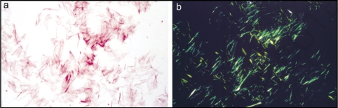
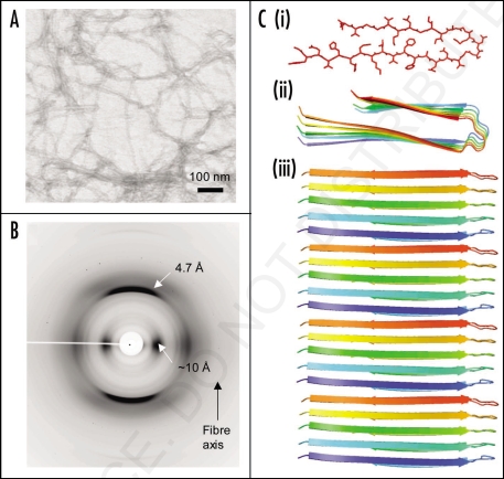
Amyloid refers to the abnormal fibrous, extracellular, proteinaceous deposits found in organs and tissues. Amyloid is insoluble and is structurally dominated by beta-sheet structure. Unlike other fibrous proteins it does not commonly have a structural, supportive or motility role but is associated with the pathology seen in a range of diseases known as the amyloidoses. These diseases include Alzheimer's, the spongiform encephalopathies and type II diabetes, all of which are progressive disorders with associated high morbidity and mortality. Not surprisingly, research into the physicochemical properties of amyloid and its formation is currently intensely pursued. In this chapter we will highlight the key scientific findings and discuss how the stability of amyloid fibrils impacts on bionanotechnology.


Figures

Figure 1
Figure 1
Isolated amyloid fibrils composed of Aα chain fragment of fibrinogen (a) stained with Congo red and visualized by light microscopy and (b) between crossed polars, showing characteristic apple-green birefringence. Figure adapted from reference .
Figure 2
Figure 2
Synthetic amyloid fibrils made from Aβ peptide (A) electron micrograph showing long, straight, unbranching fibrils. (B) X-ray fiber diffraction pattern from partially aligned amyloid fibrils showing the characteristic “cross-β” diffraction pattern. (C) The structure of the Aβ amyloid fibril interpreted from ssNMR data, showing the top view of the fiber (i and ii) with side chains (i), showing the importance of side chain packing with in the fiber and as a cartoon (ii). The side view (iii) revealing the β-strands running perpendicular to the fiber axis.
Figure 3
Figure 3
Models of mature protein fibrils based on Small-Angle X-ray scattering solution data. (A) Human alpha-synuclein fibrils and (B) human insulin fibrils. The results suggest that insulin fibrils (B) are formed of three intertwining protofibrils, whereas a-synuclein fibril (A) consist of only one protofibril. Each protofibril is assumed to consist of two intertwining protofilaments. Four and three repeating units are shown for alpha-synuclein and insulin respectively.
Figure 4
Figure 4
Amyloid-like fibers for bionanotechnology. (A) Nanowires based on the N-terminal region of the yeast prion, Sup35. Nanogold was covalently linked to the engineered cysteine residues in the protein and conjugate colloidal gold and silver particles were associated along the fibers to form wires, (B) assembly of diphenylalanine to form nanotubes that can be filled with silver to make nanowires.

References

    1. Pepys M. Pathogenesis, diagnosis and treatment of systemic amyloidosis. Phil Trans Soc Lond B. 2001;356:203–211. - PMC - PubMed
    1. Fowler DM, Koulov AV, Alory-Jost C, et al. Functional amyloid formation within mammalian tissue. PLoS Biol. 2006;4:6. - PMC - PubMed
    1. Friedrich NaK A. Zur amyloidfrage. Arch Pathol Anat Physiol Klin Med. 1859;16:50–65.
    1. Divry P, Florkin M. Sur les proprietes optiques de l'amyloid. Societe de Biologie. 1927;97:180–181. (Fre).
    1. Cohen AS, Calkins E. Electron microscopic observation on a fibrous component in amyloid of diverse origins. Nature. 1959;183:1202–1203. - PubMed

Publication types

LinkOut - more resources