

Limbal stem cell deficiency arising from systemic chemotherapy with hydroxycarbamide
- PMID: 19158571
- PMCID: PMC2670888
- DOI: 10.1097/ICO.0b013e318183a3bd
Limbal stem cell deficiency arising from systemic chemotherapy with hydroxycarbamide
Abstract
Purpose: The purpose of this study was to report a case of limbal stem cell deficiency (LSCD) after systemic chemotherapy with hydroxycarbamide.
Methods: Clinical manifestations and pathology are detailed.
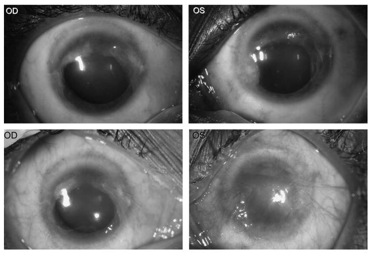
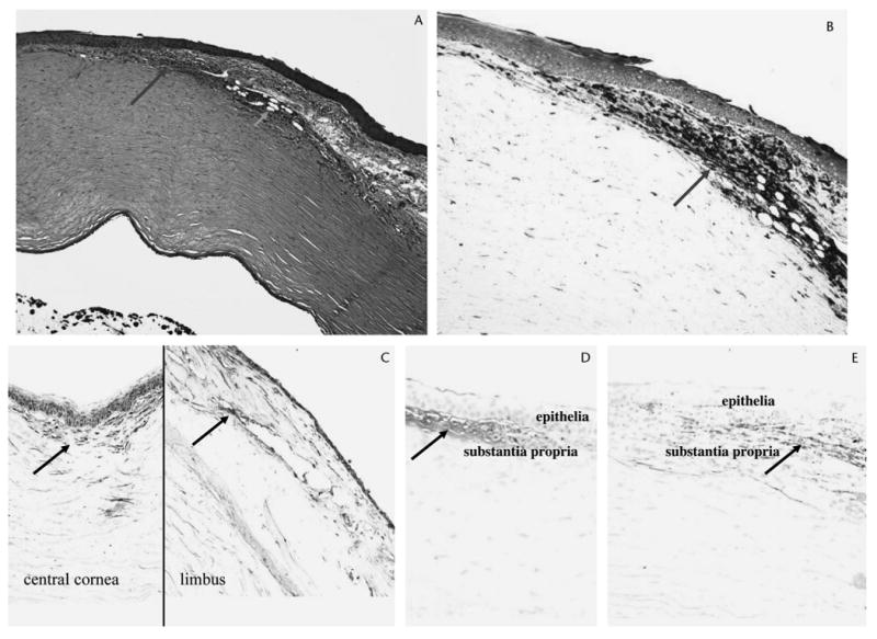
Results: We describe the case of a woman with sickle cell disease, who developed bilateral LSCD after treatment with hydroxycarbamide. Histologic examination confirmed the diagnosis of LSCD, revealing goblet cells, inflammatory cells, deposits of new collagen components, and neovascularization in the peripheral cornea. Matrix metalloproteinase-3, fibronectin, and collagen III were also detected in the lesions.
Conclusions: The systemic use of the antineoplastic drug, hydroxycarbamide, may cause severe LSCD. We recommend that a medication history, including that of cytotoxic drugs, be considered in evaluating LSCD.
Figures
Similar articles
-
The diagnosis of limbal stem cell deficiency.Ocul Surf. 2018 Jan;16(1):58-69. doi: 10.1016/j.jtos.2017.11.002. Epub 2017 Nov 4. Ocul Surf. 2018. PMID: 29113917 Free PMC article. Review.
-
Limbal stem cell transplantation: an evidence-based analysis.Ont Health Technol Assess Ser. 2008;8(7):1-58. Epub 2008 Oct 1. Ont Health Technol Assess Ser. 2008. PMID: 23074512 Free PMC article.
-
Limbal stem cell deficiency after topical mitomycin C therapy for primary acquired melanosis with atypia.Ophthalmology. 2010 Mar;117(3):431-7. doi: 10.1016/j.ophtha.2009.07.032. Epub 2010 Jan 8. Ophthalmology. 2010. PMID: 20060167
-
Treatment of partial limbal stem cell deficiency with topical interferon α-2b and retinoic acid.Br J Ophthalmol. 2016 Jul;100(7):944-948. doi: 10.1136/bjophthalmol-2015-307411. Epub 2015 Oct 27. Br J Ophthalmol. 2016. PMID: 26508779
-
Global Consensus on the Management of Limbal Stem Cell Deficiency.Cornea. 2020 Oct;39(10):1291-1302. doi: 10.1097/ICO.0000000000002358. Cornea. 2020. PMID: 32639314
Cited by
-
Global Consensus on Definition, Classification, Diagnosis, and Staging of Limbal Stem Cell Deficiency.Cornea. 2019 Mar;38(3):364-375. doi: 10.1097/ICO.0000000000001820. Cornea. 2019. PMID: 30614902 Free PMC article.
-
Disruption of Mks1 localization to the mother centriole causes cilia defects and developmental malformations in Meckel-Gruber syndrome.Dis Model Mech. 2011 Jan;4(1):43-56. doi: 10.1242/dmm.006262. Epub 2010 Nov 2. Dis Model Mech. 2011. PMID: 21045211 Free PMC article.
-
The diagnosis of limbal stem cell deficiency.Ocul Surf. 2018 Jan;16(1):58-69. doi: 10.1016/j.jtos.2017.11.002. Epub 2017 Nov 4. Ocul Surf. 2018. PMID: 29113917 Free PMC article. Review.
-
Corneal Epithelial Findings in Patients with Multiple Myeloma Treated with Antibody-Drug Conjugate Belantamab Mafodotin in the Pivotal, Randomized, DREAMM-2 Study.Ophthalmol Ther. 2020 Dec;9(4):889-911. doi: 10.1007/s40123-020-00280-8. Epub 2020 Jul 25. Ophthalmol Ther. 2020. PMID: 32712806 Free PMC article.
-
Corneal Epithelial Stem Cells-Physiology, Pathophysiology and Therapeutic Options.Cells. 2021 Sep 3;10(9):2302. doi: 10.3390/cells10092302. Cells. 2021. PMID: 34571952 Free PMC article. Review.
References
-
- Schechter AN, Rodgers GP. Sickle cell anemia—basic research reaches the clinic. N Engl J Med. 1995;332:1372–1374. - PubMed
-
- Ang LPK, Tan DTH. Ocular surface stem cells and disease: current concepts and clinical applications. Ann Acad Med Singapore. 2004;33:576–580. - PubMed
-
- Dua HS, Azuara-Blanco A. Limbal stem cells of the corneal epithelium. Surv Ophthalmol. 2000;44:415–425. - PubMed
-
- Dudney BW, Malecha MA. Limbal stem cell deficiency following topical mitomycin C treatment of conjunctival-corneal intraepithelial neoplasia. Am J Ophthalmol. 2004;137:950–951. - PubMed
Publication types
MeSH terms
Substances
Grants and funding
LinkOut - more resources
Full Text Sources
Medical
