. 2009 Mar 26;458(7237):514-8.
doi: 10.1038/nature07725. Epub 2009 Jan 21.

AIM2 recognizes cytosolic dsDNA and forms a caspase-1-activating inflammasome with ASC

Affiliations

AIM2 recognizes cytosolic dsDNA and forms a caspase-1-activating inflammasome with ASC

Veit Hornung et al. Nature. .

Abstract

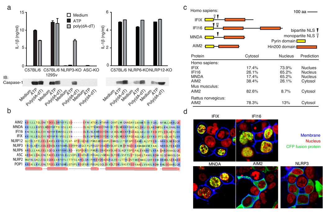
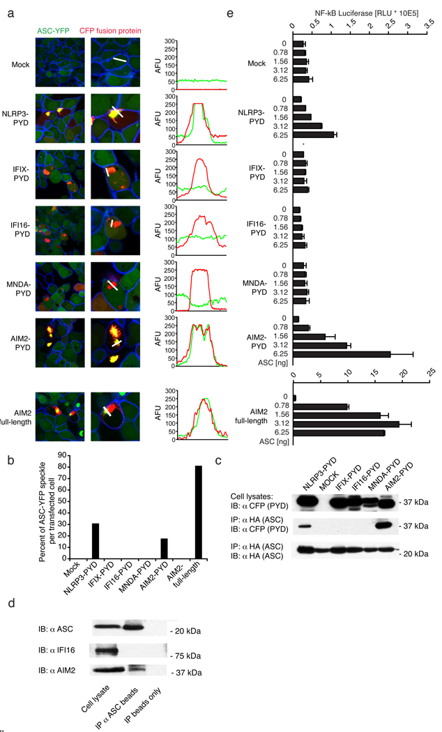
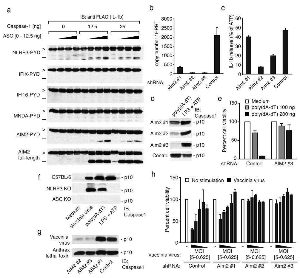
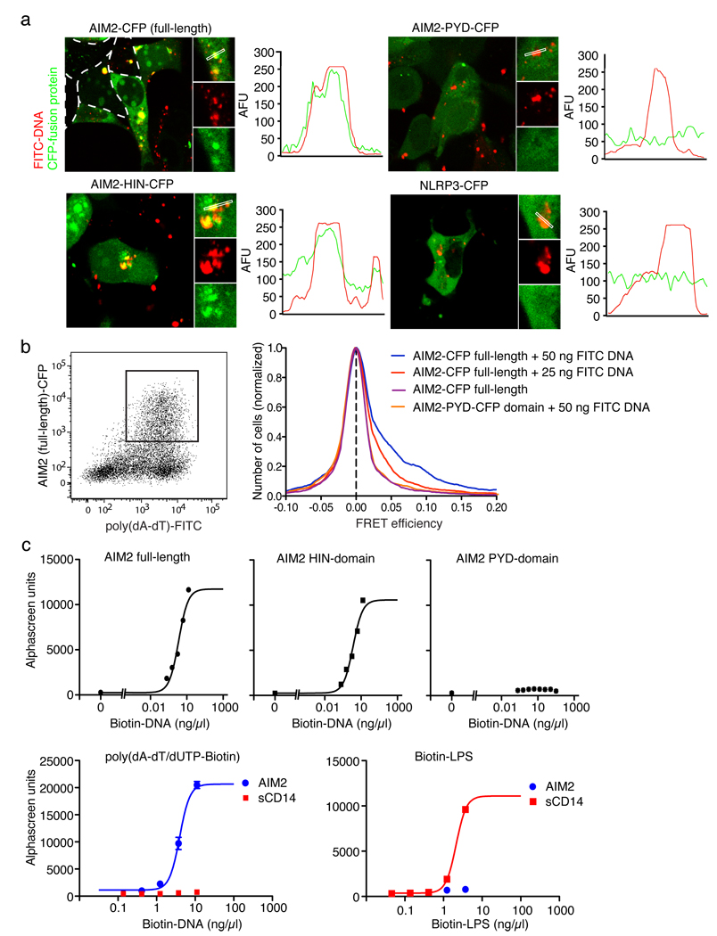
The innate immune system senses nucleic acids by germline-encoded pattern recognition receptors. RNA is sensed by Toll-like receptor members TLR3, TLR7 and TLR8, or by the RNA helicases RIG-I (also known as DDX58) and MDA-5 (IFIH1). Little is known about sensors for cytoplasmic DNA that trigger antiviral and/or inflammatory responses. The best characterized of these responses involves activation of the TANK-binding kinase (TBK1)-interferon regulatory factor 3 (IRF3) signalling axis to trigger transcriptional induction of type I interferon genes. A second, less well-defined pathway leads to the activation of an 'inflammasome' that, via caspase-1, controls the catalytic cleavage of the pro-forms of the cytokines IL1beta and IL18 (refs 6, 7). Using mouse and human cells, here we identify the PYHIN (pyrin and HIN domain-containing protein) family member absent in melanoma 2 (AIM2) as a receptor for cytosolic DNA, which regulates caspase-1. The HIN200 domain of AIM2 binds to DNA, whereas the pyrin domain (but not that of the other PYHIN family members) associates with the adaptor molecule ASC (apoptosis-associated speck-like protein containing a caspase activation and recruitment domain) to activate both NF-kappaB and caspase-1. Knockdown of Aim2 abrogates caspase-1 activation in response to cytoplasmic double-stranded DNA and the double-stranded DNA vaccinia virus. Collectively, these observations identify AIM2 as a new receptor for cytoplasmic DNA, which forms an inflammasome with the ligand and ASC to activate caspase-1.


Figures

Figure 1
Figure 1. poly(dA-dT)-induced inflammasome activation
a, LPS-primed macrophages from wild type or inflammasome deficient mice were stimulated as indicated and supernatants examined for IL-1β by ELISA or for cleaved caspase-1 by immunoblot. b, A multiple sequence alignment of human PYHIN and select NLR PYD domains, c, Domain structures of human PYHINs, with predicted nuclear localization signals and subcellular localizations (lower panel). d, Subcellular localization of CFP-tagged IFIX, IFI16, MNDA, AIM2 or NLRP3 (green) in 293T cells. Fluorescent choleratoxin stained membranes (blue) and DRAQ5-stained nuclei (red). Data from one experiment of three is shown (a, d).
Figure 2
Figure 2. AIM2 interacts with ASC
a, ASC-YFP-expressing cells (green) were transfected as indicated and imaged by confocal microscopy. Fluorescence intensities of green (YFP-ASC) and red (PYD-CFP) channels were quantified along white lines and b, the percentage of ASC-YFP speckles shown. c, 293T cells transfected with HA-ASC and CFP-tagged constructs as in (a) were immunoprecipitated (IP) with anti-HA and immunoblotted (WB) as indicated (lower panel). d, ASC was immunoprecipitated from Sendai virus primed THP-1 cells and ASC, IFI16 and AIM2 examined by immunoblotting. The band above the AIM2 band in the ASC immunoprecipitation corresponds to the heavy chain of the anti-ASC antibody e, NF-κB luciferase reporter gene activity was measured upon transfection with the indicated plasmids. Data of one representative experiment out of three (a, b, c and d) or two (e) are depicted.
Figure 3
Figure 3. AIM2 is required for poly(dA-dT)- and vaccinia virus-triggered inflammasome activation
a, 293T cells were transfected as indicated and cell lysates immunoblotted for pro-IL-1β (>) and cleaved IL-1β (−). b, B6-MCLs were transduced with lentiviral vectors encoding shRNAs as indicated and AIM2/HPRT1 measured by QPCR. c, LPS-primed cells as in (b) were stimulated as indicated for 6 hrs and IL-1β in supernatants measured by ELISA. The poly(dA-dT) triggered IL-1β release was normalized to the ATP induced IL-1β levels. Absolute values for ATP-triggered IL-1β release were 1790, 2078, 2676 and 1119 pg/ml (AIM2#1, AIM2#2, AIM2#3 and Control) respectively. d, Macrophages as in (b) were treated as indicated and cleavage of caspase-1 measured by immunoblotting after 6h. e, Macrophages transduced with shRNA were transfected as indicated and cells counted 24h later. f, Macrophages from the indicated strains were treated as indicated and cleavage of caspase-1 measured after 6h. g, NLRP3-deficient shRNA expressing macrophages as in (b) were infected as indicated and assessed for cleavage of caspase-1 after 6h. h, Cells as in (b) were infected as indicated and cell survival measured by calcein AM staining 24 h later. One representative experiment out of three (a, d, e, f, g and h) four (c) or five (b) is depicted.
Figure 4
Figure 4. AIM2 binds dsDNA via its HIN domain
a, 293T cells were transfected with CFP-tagged AIM2 or NLRP3 (red) as indicated together with fluorescein labeled poly(dA-dT) (FITC-DNA, green) and imaged by confocal microscopy. Fluorescence intensities of the green (FITC-DNA) and red (CFP fusion protein) channels were quantified along selected lines. b, 293T cells were transfected as indicated with FITC-DNA or unlabeled DNA. Cells were analyzed by flow cytometry after 24 h and CFP and FITC positive cells were gated (left panel) and analyzed for FRET efficiency on a cell-by-cell basis. Calculated FRET efficiency histograms are shown (right panel). c, AlphaScreen assessment of AIM2 full length, AIM2-HIN domain or AIM2-PYD domain binding to poly(dA-dT)/Biotin-dUTP (Biotin-DNA) (upper panel) and AIM2 or sCD14 binding to Biotin-DNA or biotin LPS (lower panel). Representative data from 2 (d) or three (a, b, c) independent experiments are shown.

Similar articles

Cited by

References

    1. Meylan E, Tschopp J, Karin M. Intracellular pattern recognition receptors in the host response. Nature. 2006;442(7098):39. - PubMed
    1. Ishii KJ, et al. A Toll-like receptor-independent antiviral response induced by double-stranded B-form DNA. Nat Immunol. 2006;7(1):40. - PubMed
    1. Stetson DB, Medzhitov R. Recognition of cytosolic DNA activates an IRF3-dependent innate immune response. Immunity. 2006;24(1):93. - PubMed
    1. Takaoka A, et al. DAI (DLM-1/ZBP1) is a cytosolic DNA sensor and an activator of innate immune response. Nature. 2007;448(7152):501. - PubMed
    1. Ishii KJ, et al. TANK-binding kinase-1 delineates innate and adaptive immune responses to DNA vaccines. Nature. 2008;451(7179):725. - PubMed

Publication types

MeSH terms