. 2009;4(1):e4253.
doi: 10.1371/journal.pone.0004253. Epub 2009 Jan 22.

Impaired nuclear Nrf2 translocation undermines the oxidative stress response in Friedreich ataxia

Affiliations

Impaired nuclear Nrf2 translocation undermines the oxidative stress response in Friedreich ataxia

Vincent Paupe et al. PLoS One. 2009.

Abstract

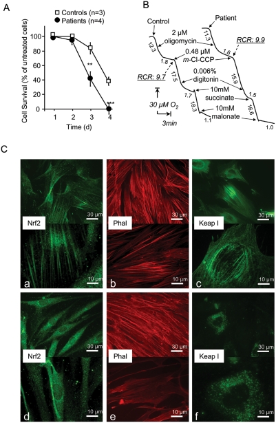
Background: Friedreich ataxia originates from a decrease in mitochondrial frataxin, which causes the death of a subset of neurons. The biochemical hallmarks of the disease include low activity of the iron sulfur cluster-containing proteins (ISP) and impairment of antioxidant defense mechanisms that may play a major role in disease progression.

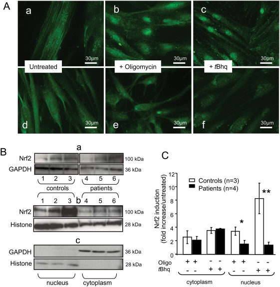
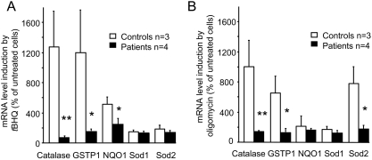
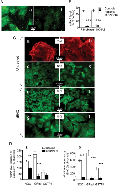
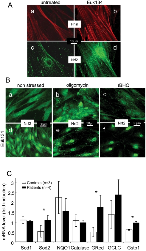
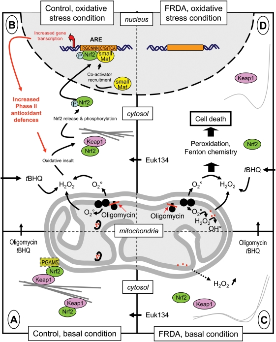
Methodology/principal findings: We thus investigated signaling pathways involved in antioxidant defense mechanisms. We showed that cultured fibroblasts from patients with Friedreich ataxia exhibited hypersensitivity to oxidative insults because of an impairment in the Nrf2 signaling pathway, which led to faulty induction of antioxidant enzymes. This impairment originated from previously reported actin remodeling by hydrogen peroxide.

Conclusions/significance: Thus, the defective machinery for ISP synthesis by causing mitochondrial iron dysmetabolism increases hydrogen peroxide production that accounts for the increased susceptibility to oxidative stress.


Conflict of interest statement

Competing Interests: The authors have declared that no competing interests exist.

Figures

Figure 1
Figure 1. Effect of frataxin depletion on oxidative stress resistance, oxidative properties, and actin-Nrf2 signaling pathway status in cultured skin fibroblasts.
A. Sensitivity of control and patient cells to oligomycin (30 µM for 4 days). Cells were harvested at 24-hour intervals and live cells were counted. The proportion of surviving cells was significantly different between controls and patients (ANOVA p<0.01). Significant differences were noted (**p<0.01 and ***p<0.001). Values are means±1 SEM. Open and dark symbols represent control and patient cells, respectively. Filled squares represent patients. B. Effect of frataxin depletion on cell oxidative properties. Under basal conditions, respiration rates were similar between fibroblasts from controls and patients. Adding oligomycin caused greater than 80% inhibition in both cell types. Uncoupled respiration measured in the presence of m-Cl-CCP (carbonyl cyanide m-chlorophenylhydrazone) decreased progressively when digitonin was added to induce cell permeabilization. Malonate-sensitive mitochondrial succinate oxidation was not different between control and patient cells. Numbers along the traces are nmol/min/mg protein. C. Nrf2/Keap1 localization in patient and control fibroblasts. Labeling of Nrf2, actin (Phal), and Keap1 proteins in control (a, b, c) and patient (e, f, g) fibroblasts, showing disorganization of the actin network and abnormal location of Nrf2 and Keap1 in patient fibroblasts. Experimental procedures are described in the methods section.
Figure 2
Figure 2. Nrf2 location and amount in control and FRDA patient fibroblasts under basal and oxidative stress conditions.
A. Nrf2 localization in control (a, b, c) and patient (d, e, f) fibroblasts under basal conditions (a, d) or after treatment with oligomycin (b, e) or tBHQ (c, f). Nuclear Nrf2 translocation occurred in control cells, but not patient cells, after oligomycin or tBHQ treatment. B. Western blots of cytoplasmic (a; 40 µg/lane protein) and nuclear (b; 60 µg/lane protein) fractions from control (lanes 1–3) and patient (lanes 4–6) fibroblasts under basal conditions (lane 1, 4), after oligomycin treatment (lane 2, 5), or after tBHQ treatment (lane 3, 6). The specificity of the 100 kDa band was confirmed using two antibodies (H-300 and C-20). Fraction purity (d) was assessed by labeling with GAPDH (cytoplasm) and Histone H1 antibody (nuclei). C. Nrf2 content relative to GAPDH and Histone H1 contents in cytoplasmic and nuclear fractions of control and patient fibroblasts. Asterisks denote significant differences (*p<0.05 and **p<0.01). Values are means±1 SEM. Experimental procedures are described in the methods section.
Figure 3
Figure 3. Induction of phase II antioxidants mediated by Nrf2 in control and patient fibroblasts treated with tBHQ or oligomycin.
Transcription induced by tBHQ (A) or oligomycin (B). Significant differences were noted (*p<0.05 and **p<0.01). Values are means±1 SEM. Experimental procedures are described in the methods section.
Figure 4
Figure 4. Frataxin-depleted SKNAS cells.
A. Anti-frataxin antibody produced strong mitochondrial labeling in control SKNAS cells (a) and severely reduced labeling in SKNAS cells treated with frataxin-targeted shRNA (shRNAFra SKNAS cells) (b). B. Residual frataxin mRNA in patient fibroblasts and shRNAFra SKNAS cells compared to control cells. C. Actin and Nrf2 in control (a, c, e, g) and shRNAFra (b, d, f, h) SKNAS cells under basal conditions (a, b, c, d) or after tBHQ treatment (e, f, g, h). Actin staining with phalloidin (Phal) shows disorganization of the actin stress fibers in shRNAFra SKNAS cells (a) compared to control SKNAS cells (b), in keeping with the results in fibroblasts. Nrf2 labeling produced similar abundant staining of the cytoplasm of both control SKNAS cells (c) and shRNAFra SKNAS cells (d). Nuclear translocation of Nrf2 occurs in control SKNAS cells (e, g) but not in shRNAFra SKNAS cells. D. Induction of Phase II antioxidants in SKNAS cells using oligomycin (a) or tBHQ (b). Significant differences were noted (**p<0.01 and *** p<0.001). Values are means±1 SEM. Experimental procedures are described in the methods section.
Figure 5
Figure 5. Effect of Euk134 on Nrf2 localization under basal conditions and during oxidative stress in fibroblasts from patients with Friedreich ataxia.
A. Staining of actin (a,b) and Nrf2 (c,d) in untreated (a, c) and treated (200 µM Euk134 for 24 h) (b, d) patient fibroblasts. Euk134 restored the normal actin network organization and the location of Nrf2 on the actin bundles. B. Patient fibroblasts under basal conditions (a, d), with oligomycin treatment (b, e), or with tBHQ treatment (c, f) after 24 hours pretreatment with 200 µM Euk134 (d, e, f) or without pretreatment (a, b, c). In pre-treated cells, oligomycin and tBHQ induced massive nuclear translocation of Nrf2. C. Induction of Phase II antioxidants in control and patient fibroblasts treated with oligomycin. Treatments and experimental procedures are described in the methods section. Significant differences were noted (*p<0.05). Values are means±1 SEM.
Figure 6
Figure 6. Diagram of the actin-Nrf2 signaling pathway in control and patient cells under basal conditions and during oxidative stress.
Under basal conditions, the Nrf2-Keap1 complex, or a sub-pool of it attached to the mitochondrial outer membrane by the PGAM5 protein, is bound to the actin stress filament network of control cells. B. In frataxin-depleted cells, characterized by abnormal iron handling, the actin-Nrf2 signaling pathway is profoundly altered by the need to cope with elevated H2O2 levels. Removing H2O2 with the catalase mimetic Euk134 corrects these alterations. C. Treating control cells with oligomycin or tBHQ results in major oxidative stress that destabilizes the actin-Nrf2-Keap1 complex, leading to Nrf2 release and phosphorylation. Nuclear translocation of Nrf2 results in the recruitment of the co-activator(s) needed for Phase II antioxidant transcription. D. In frataxin-depleted cells, the oxidative insult induced by oligomycin (endogenous) or tBHQ (exogenous) cannot be counterbalanced by the induction of Phase II antioxidants, so that the cells are extremely sensitive to oxidation. Again, Euk134 treatment restores the actin-Nrf2 signaling pathway, allowing transcription of Phase II antioxidants.

References

    1. Tzagoloff A. Mitochondria. New York: Plenum Press; 1982.
    1. Rustin P. Mitochondria, from cell death to proliferation. Nat Genet. 2002;30:352–3. - PubMed
    1. Han D, Williams E, Cadenas E. Mitochondrial respiratory chain-dependent generation of superoxide anion and its release into the intermembrane space. Biochem J. 2001;353:411–6. - PMC - PubMed
    1. Koehler CM, Beverly KN, Leverich EP. Redox pathways of the mitochondrion. Antioxid Redox Signal. 2006;8:813–22. - PubMed
    1. Droge W. Free radicals in the physiological control of cell function. Physiol Rev. 2002;82:47–95. - PubMed

Publication types