. 2009:15:168-80.
Epub 2009 Jan 21.

Overexpression of CERKL, a gene responsible for retinitis pigmentosa in humans, protects cells from apoptosis induced by oxidative stress

Affiliations

Overexpression of CERKL, a gene responsible for retinitis pigmentosa in humans, protects cells from apoptosis induced by oxidative stress

Miquel Tuson et al. Mol Vis. 2009.

Abstract

Purpose: Retinitis pigmentosa (RP), a retinal neurodegenerative disorder characterized by apoptosis of photoreceptor cells, is caused by mutations in many different genes. We analyzed the RP gene ceramide kinase-like (CERKL) to determine CERKL function and contribution to pathogenesis.

Methods: RT-PCR was performed to characterize CERKL expression in many human adult and fetal tissues, including retina. We analyzed the protein subcellular localization by confocal microscopy and further verified it by sucrose gradients. We performed lipid kinase activity assays. And finally, we studied the effects on cell apoptosis after CERKL overexpression in transiently transfected cultured cells by propidium iodide staining and poly-(ADP-ribose)-polymerase (PARP) caspase-dependent cleavage.

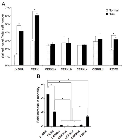
Results: CERKL transcripts underwent alternative splicing. In the human retina, four different CERKL isoforms of 532, 558, 419, and 463 amino acids were expressed. CERKL proteins were mainly localized in the endoplasmic reticulum and Golgi compartments, but they also shifted localization to nuclei and nucleoli. We also found that CERKL prevented cells from entering apoptosis induced by oxidative-stress conditions.

Conclusions: CERKL remains a unique orphan lipid kinase in that no candidate substrate has been identified after intense research. The dynamic localization of CERKL suggests multiple sites of action. Remarkably, CERKL (but not the RP R257X mutant) exerts a protective role in cells against oxidative stress, consistent with RP mutations impairing the normal protein function in photoreceptors and thus tilting the balance toward apoptosis. These results provide valuable insights into the molecular mechanisms causing retinal degeneration.


Figures

Figure 1
Figure 1
CERKL isoform structure. A: The diagram shows the genomic exon-intron structure of the CERKL gene with the initiation and stop codons. The position of the nonsense mutation (R257X) identified in RP families as well as the splicing junctions resulting in isoforms a, b, c, and d are indicated. Asterisks indicate the position of the rare GC-splicing donor sites. B: The mature CERKL mRNA isoforms, indicating the relative position of the encoded DAG kinase and putative PH domains, are depicted. Note that exon 4b (isoform b, in gray) is located well within the DAG kinase domain, while isoforms c and d lack this domain. Grey small double arrows indicate the position of the primers used for specific isoform RT–PCR analyses (see Methods). The figure is drawn to scale except for the introns depicted with a broken line.
Figure 2
Figure 2
RT–PCR analysis of specific CERKL isoforms in several human tissues. For each tissue, lane 1 shows amplification of isoforms a (fast migrating band) and b (slow migrating band), whereas lanes 2 and 3 show the amplified product of isoform c and d, respectively. In the second gel, “f” represents fetal tissue. Marker band sizes are indicated in base pairs (bp). The additional faint bands in lane number 1 correspond to heteroduplexes of the PCR products.
Figure 3
Figure 3
CERKL kinase activity assays. A: The autoradiography of one out of many thin layer chromatographies (TLC) from in vitro assays is shown. Lipid micelles and heat-inactivated cell lysates from 661W (murine photoreceptor-derived cell line) cells were used as substrates, whereas protein lysates from COS-7 cells transfected with either empty vector (pcDNA), CERKL isoform a (532 aa), CERKL isoform b (558 aa) or ceramide kinase CERK (positive control) expressing constructs were the source of the enzymatic activity. B: Autoradiography of a TLC from in vivo assays is shown. Cultured 661W cells were transfected with either pGFP (empty vector), or CERKLa-GFP, CERKLb-GFP, or CERK-GFP (positive control), selected by FACS and grown in a medium supplemented with 32P-orthophosphate (see Material and Methods for details on the protocols). C1P represents Ceramide-1-phosphate. The images are representative of many different assays, with several experimental parameters, such as the type of cell line and the CERKL isoforms overexpressed, changed. The results of the assays were negative under all the conditions tested.
Figure 4
Figure 4
CERKL-HA (isoform a, 532 amino acids) shows a dynamic subcellular localization in COS-7 transfected cells. A: Several cells per field showed uniform distribution of CERKL in the cytosol and the nucleus, with clear exclusion from the nucleoli. B: A similar pattern but with strong accumulation in the nucleoli (nuclei, counter-stained with DAPI, appear in blue). C and D: In most cells, CERKL was absent from the nucleus and instead accumulated in clusters, preferentially in the perinuclear region (A). These two patterns were cell-specific and could be observed in the same field (D). E: CERKL localized to several membranous subcellular compartments, mainly ER and Golgi. The markers used were calnexin for ER, GM130 for Golgi, EEA1 for endosomes and MitoTracker for mitochondria. F: R257X truncated CERKL localized preferentially in the nuclei, although it was also detected in the ER. Arrows highlight the relevant CERKL localizations, as immunodetected with an anti-HA monoclonal antibody. Scale bar corresponds to 10 μm. The same results were obtained for all the CERKL isoforms, irrespective of the epitope used, HA or GFP (data not shown). Abbreviations: endoplasmic reticulum (ER), endosomes (End), mitochondria (Mit).
Figure 5
Figure 5
CERKL colocalizes with Trans-Golgi, Golgi and ER markers. HeLa cells transfected with CERKLa-HA were submitted to subcellular fractionation by sucrose gradient. The strongest signal of CERKLa coincides with the Trans-Golgi TGN38 marker, but it is also localized in the same fractions as the Golgi GM130 marker and extends to the ER PDI marker fractions. The multiple banded pattern observed in the TGN38 immunodetection has been previously reported as this protein undergoes complex post-translational modification events. CERKLa-HA was immunodetected with a monoclonal anti-HA antibody. The lanes are numbered according to the sucrose fractions as collected from 1 (bottom, with higher sucrose concentration) to 10 (top, lower sucrose concentration). Abbreviations: total protein lysate (L); supernatant after pelleting the membranes by centrifugation at 100,000x g (S).
Figure 6
Figure 6
Cells overexpressing CERKL showed no mortality increase in a comparison of normal and oxidative stress conditions. A: The histogram shows the mortality (depicted as percentage) of pyknotic nuclei of COS-7 cells. Cells were transfected with either the wild-type CERKL isoforms (named CERKLa to CERKLd), the truncated RP form (R257X), CERK or the empty vector (negative control) and grown in either normal conditions (dotted bars) or treated for 4 h with 200 μM H2O2 (solid bars). There was no increase in mortality when the cells were transfected with any of the wt CERKL isoforms. In contrast, there was an increase in mortality in cells transfected with, either the empty vector, a CERK (ceramide kinase) construct or the R257X CERKL mutant. Statistical significance is shown by an asterisk (Mann–Whitney test, p<0.05). B: The histogram shows the mortality fold-increase of H2O2-treated versus untreated cells for each construct. Statistically significant differences are shown by asterisks (Mann–Whitney test, p<0.05). More than 500 cells were counted for each construct and condition, in 3 independent replicates.
Figure 7
Figure 7
Overexpression of CERKL protects HeLa cells from apoptosis caused by oxidative stress. A: The western blot of the PARP apoptosis-dependent cleavage was obtained and immunodetected 24 h after treatment with several oxidative reagents, as indicated. The PARP precursor protein size is 116 kDa, whereas the proteolytic product after caspase-3 cleavage is 85 kDa. Cells were transfected with either the CERKLa (532aa, encompassing the kinasic domain) or the empty vector pcDNA3. Immunodetection of tubulin was used for normalization. This is one image of several similar replicates. The transfection efficiency was comparable as assessed by western immunodetection (data not shown). B and C: Quantification of the PARP-cleaved peptide with respect to total PARP immunodetected protein (both precursor plus peptide) in empty vector (red bars) versus CERKLa transfected cells (solid bars) under the different treatments. Basal apoptosis in empty vector untreated cells was arbitrarily considered 100%. CERKL protection against apoptosis was clearly detected after 12 h treatment with 300 μM H2O2 compared with the empty-vector transfected cells, whereas in the other oxidative conditions this protective effect was not significant (B). This protective effect of CERKL against apoptosis induced by 300 μM H2O2 was much more pronounced after 24 h treatment (C). D: The histogram shows the quantification of the PARP-cleaved peptide with respect to total PARP immunodetected protein (both precursor plus peptide) in empty vector-transfected cells (red bars) versus cells transfected with either CERKLa (solid bars), the R257X CERKLa mutant (white bars) or the R257X CERKLb mutant (blue bars). Cells were grown under normal conditions or treated with different concentrations of H2O2. Basal apoptosis in empty vector untreated cells was arbitrarily considered 100%. The protective effect is clearly detected for the full-length protein but not for the truncated mutants. NT-untreated cells; 300-cells treated with 300 μM H2O2; 400-cells treated with 400 μM H2O2; FBS-cells grown in medium deprived of fetal bovine serum; SNP-cells grown in medium supplemented with 0.3 mM sodium nitroprusside; R257Xa- cells transfected with the construct bearing the R257X mutation in the CERKLa sequence background; R257Xb-cells transfected with the construct bearing the R257X mutation in the CERKLb sequence background. At least 3 independent experiments were used for replication. Statistical significance is indicated by an asterisk (Mann–Whitney test, p<0.05).

Similar articles

Cited by

References

    1. Bayés M, Goldaracena B, Martínez-Mir A, Iragui-Madoz MI, Solans T, Chivelet P, Bussaglia E, Ramos-Arroyo MA, Baiget M, Vilageliu L, Balcells S, Gonzàlez-Duarte R, Grinberg D. A new autosomal recessive retinitis pigmentosa locus maps on chromosome 2q31-q33. J Med Genet. 1998;35:141–5. - PMC - PubMed
    1. Tuson M, Marfany G, Gonzalez-Duarte R. Mutation of CERKL, a novel human ceramide kinase gene, causes autosomal recessive retinitis pigmentosa (RP26). Am J Hum Genet. 2004;74:128–38. - PMC - PubMed
    1. Auslender N, Sharon D, Abbasi AH, Garzozi HJ, Banin E, Ben-Yosef T. A common founder mutation of CERKL underlies autosomal recessive retinal degeneration with early macular involvement among Yemenite Jews. Invest Ophthalmol Vis Sci. 2007;48:5431–8. - PubMed
    1. Hannun YA, Obeid LM. The ceramide-centric universe of lipid-mediated cell regulation: stress encounters of the lipid kind. J Biol Chem. 2002;277:25847–50. - PubMed
    1. Hannun YA, Obeid LM. Principles of bioactive lipid signalling: lessons from sphingolipids. Nat Rev Mol Cell Biol. 2008;9:139–50. - PubMed

Publication types

MeSH terms