. 2009 Jan;5(1):e1000284.
doi: 10.1371/journal.ppat.1000284. Epub 2008 Jan 30.

Glutathione provides a source of cysteine essential for intracellular multiplication of Francisella tularensis

Affiliations

Glutathione provides a source of cysteine essential for intracellular multiplication of Francisella tularensis

Khaled Alkhuder et al. PLoS Pathog. 2009 Jan.

Abstract

Francisella tularensis is a highly infectious bacterium causing the zoonotic disease tularemia. Its ability to multiply and survive in macrophages is critical for its virulence. By screening a bank of HimarFT transposon mutants of the F. tularensis live vaccine strain (LVS) to isolate intracellular growth-deficient mutants, we selected one mutant in a gene encoding a putative gamma-glutamyl transpeptidase (GGT). This gene (FTL_0766) was hence designated ggt. The mutant strain showed impaired intracellular multiplication and was strongly attenuated for virulence in mice. Here we present evidence that the GGT activity of F. tularensis allows utilization of glutathione (GSH, gamma-glutamyl-cysteinyl-glycine) and gamma-glutamyl-cysteine dipeptide as cysteine sources to ensure intracellular growth. This is the first demonstration of the essential role of a nutrient acquisition system in the intracellular multiplication of F. tularensis. GSH is the most abundant source of cysteine in the host cytosol. Thus, the capacity this intracellular bacterial pathogen has evolved to utilize the available GSH, as a source of cysteine in the host cytosol, constitutes a paradigm of bacteria-host adaptation.


Conflict of interest statement

The authors have declared that no competing interests exist.

Figures

Figure 1
Figure 1. Intracellular replication of LVS, ggt, and ggt-complemented strains.
Replication of LVS, ggt, and ggt-complemented strains, inside J774 murine macrophage-like cells (A); RAW macrophages (B); mouse bone marrow-derived macrophages (BMM) from BALB/c mice (C); and THP1 human macrophages (D). Results are shown as the average log10 CFU/well±standard deviation. Strains are denoted as follows: LVS-pKK214 (◆), ggt -pKK214 (□), and ggt-pKK-ggt (■).
Figure 2
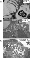
Figure 2. Intracellular electron microscopy.
Transmission electron micrographs of thin sections of RAW macrophages infected by LVS or ggt, at T0 and after 24 h infection. At T0, F. tularensis initially resides in a spacious vacuole at the periphery of the cell (A): LVS. The arrows point to regions where the phagosomal membrane surrounding the bacteria is visible. At 24 h, F. tularensis LVS (B) or ggt (C) localized in the cytoplasm are surrounded by an electron-lucent space. (B,C) Illustrate cytosolic multiplication of LVS and the ggt mutant, respectively. The bar to the bottom left corresponds to 0.2 µm (A) or 1 µm (B,C).
Figure 3
Figure 3. GGT is important for virulence in mice.
Survival of BALB/c mice after i.p. infection with LVS, ggt, or ggt-complemented (ggt_C) bacteria. Groups of five mice were infected with the number of bacteria (log10) shown next to the curves and followed for 9 days.
Figure 4
Figure 4. Killing of LVS and mutants by non-immune human serum.
Bacteria were incubated with increasing concentrations of serum, and samples were collected after one hour incubation at 37°C. The number of viable bacteria was determined by plating onto chocolate agar plates.
Figure 5
Figure 5. Cysteine is required for F. tularensis growth.
(A) Growth of LVS and the ggt mutant in chemically defined medium (CMD) supplemented with various sources of cysteine. The OD600 of the cultures were monitored during 12 h of growth at 37°C with agitation in complete CDM (CDM+) or in CDM lacking cysteine (CDM−) and supplemented with either γ-Glu-Cys or GSH. (B) Replication of LVS and ggt mutant inside J774 cells in the presence or absence of cysteine 5 mM (left panel); or in the presence or absence of γ-Glu-cys 1 mM (right panel).
Figure 6
Figure 6. Functional complementation of E. coli ggt with ggt of F. tularensis.
E. coli auxotrophic strains SH794 (thr leu ggt) and SH795 (thr leu) containing pKK214 or pKK-ggt (encoding F. tularensis GGT), grown on M9 minimal agar plates supplemented with threonine and either leucine (Leu), γ-glytamyl-leucine (γGluLeu), or no leucine source (none).
Figure 7
Figure 7. Metabolic labeling.
LVS and ggt mutant bacteria were grown in CDM devoid of cysteine, supplemented with either GSH (lanes LVS+GSH, ggt+GSH) or cysteine (lanes LVS+Cys, ggt+Cys). Radiolabeled 35S-GSH was added to each culture and the suspensions were incubated for 8 h at 37°C with agitation. Bacterial pellets were resuspended in SDS-PAGE loading buffer, and 5 µl of each fraction were loaded onto 10%-SDS-polyacrilamide gels. Each well corresponds to ca. 1.8×10⁸ bacteria for LVS+GSH, LVS+Cys, and ggt+Cys; and to 0.6×10⁸ bacteria for ggt+GSH. After electrophoresis, gels were vacuum-dried and scanned with a Molecular Dynamics Phosphorimager. The autogradiograph shown in (A) corresponds to 12 h exposure. As control, a second gel loaded with the same fractions was Coomassie-blue-stained (B).
Figure 8
Figure 8. A model of utilization of GSH and γ-Glu-Cys by cytosolic F. tularensis.
F. tularensis requires cysteine for growth. It can utilize GSH (and its oxidized form GSSG), γ-Glu-Cys, cysteine, and cystine as sources of cysteine. In eukaryotic host cells, the intracellular concentration of GSH depends on the availability and transport of cystine (oxidized form of cysteine) and cysteine. Cytosolic GSH, the most abundant thiol-containing compound (present in the mM range), and γ-Glu-Cys serve as sources of cysteine for intracellular growth of F. tularensis. In wild-type bacteria, GSH and γ-Glu-Cys are processed by GGT to produce γ-Glu and Cys-Gly or γ-Glu and Cys, respectively. The Cys-Gly dipeptide is further processed by other amino-acid peptidases to produce free cysteine and glycine. GGT-negative bacteria are unable to process these compounds, and the available concentration of free cysteine is too low to promote bacterial growth.

References

    1. Titball RW, Sjostedt A, Pavelka MS, Jr, Nano FE. Biosafety and selectable markers. Ann N Y Acad Sci. 2007;1105:405–417. - PubMed
    1. Ellis J, Oyston PC, Green M, Titball RW. Tularemia. Clin Microbiol Rev. 2002;15:631–646. - PMC - PubMed
    1. Sjostedt A. Tularemia: history, epidemiology, pathogen physiology, and clinical manifestations. Ann N Y Acad Sci. 2007;1105:1–29. - PubMed
    1. Barker JR, Klose KE. Molecular and genetic basis of pathogenesis in Francisella tularensis. Ann N Y Acad Sci. 2007;1105:138–159. - PubMed
    1. Brotcke A, Weiss DS, Kim CC, Chain P, Malfatti S, et al. Identification of MglA-regulated genes reveals novel virulence factors in Francisella tularensis. Infect Immun. 2006;74:6642–6655. - PMC - PubMed

Publication types

MeSH terms

LinkOut - more resources