Aluminum nanoparticles as substrates for metal-enhanced fluorescence in the ultraviolet for the label-free detection of biomolecules
- PMID: 19159327
- PMCID: PMC2729167
- DOI: 10.1021/ac802118s
Aluminum nanoparticles as substrates for metal-enhanced fluorescence in the ultraviolet for the label-free detection of biomolecules
Abstract
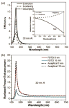
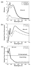
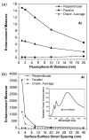
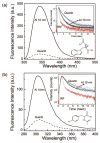
We use finite-difference time-domain calculations to show that aluminum nanoparticles are efficient substrates for metal-enhanced fluorescence (MEF) in the ultraviolet (UV) for the label-free detection of biomolecules. The radiated power enhancement of the fluorophores in proximity to aluminum nanoparticles is strongly dependent on the nanoparticle size, fluorophore-nanoparticle spacing, and fluorophore orientation. Additionally, the enhancement is dramatically increased when the fluorophore is between two aluminum nanoparticles of a dimer. Finally, we present experimental evidence that functionalized forms of amino acids tryptophan and tyrosine exhibit MEF when spin-coated onto aluminum nanostructures.
Figures


Similar articles
-
Aluminum nanostructured films as substrates for enhanced fluorescence in the ultraviolet-blue spectral region.Anal Chem. 2007 Sep 1;79(17):6480-7. doi: 10.1021/ac071363l. Epub 2007 Aug 8. Anal Chem. 2007. PMID: 17685553 Free PMC article.
-
Deep-UV surface-enhanced resonance Raman scattering of adenine on aluminum nanoparticle arrays.J Am Chem Soc. 2012 Feb 1;134(4):1966-9. doi: 10.1021/ja210446w. Epub 2012 Jan 19. J Am Chem Soc. 2012. PMID: 22239484
-
Enhanced fluorescence of proteins and label-free bioassays using aluminum nanostructures.Anal Chem. 2009 Aug 1;81(15):6049-54. doi: 10.1021/ac900263k. Anal Chem. 2009. PMID: 19594133 Free PMC article.
-
Advances in three dimensional metal enhanced fluorescence based biosensors using metal nanomaterial and nano-patterned surfaces.Biotechnol J. 2024 Jan;19(1):e2300519. doi: 10.1002/biot.202300519. Epub 2023 Dec 5. Biotechnol J. 2024. PMID: 37997672 Review.
-
Wavelength-Dependent Metal-Enhanced Fluorescence Biosensors via Resonance Energy Transfer Modulation.Biosensors (Basel). 2023 Mar 13;13(3):376. doi: 10.3390/bios13030376. Biosensors (Basel). 2023. PMID: 36979588 Free PMC article. Review.
Cited by
-
The use of aluminum nanostructures as platforms for metal enhanced fluorescence of the intrinsic emission of biomolecules in the ultra-violet.Proc SPIE Int Soc Opt Eng. 2010 Feb;7577:75770O. doi: 10.1117/12.841449. Proc SPIE Int Soc Opt Eng. 2010. PMID: 20706552 Free PMC article.
-
Metal-enhanced intrinsic fluorescence of nucleic acids using platinum nanostructured substrates.Chem Phys Lett. 2012 Oct 1;548:45-50. doi: 10.1016/j.cplett.2012.08.020. Chem Phys Lett. 2012. PMID: 23002289 Free PMC article.
-
Programmable and reversible plasmon mode engineering.Proc Natl Acad Sci U S A. 2016 Dec 13;113(50):14201-14206. doi: 10.1073/pnas.1615281113. Epub 2016 Nov 28. Proc Natl Acad Sci U S A. 2016. PMID: 27911819 Free PMC article.
-
Aluminum plasmonic nanoshielding in ultraviolet inactivation of bacteria.Sci Rep. 2017 Aug 22;7(1):9026. doi: 10.1038/s41598-017-08593-8. Sci Rep. 2017. PMID: 28831133 Free PMC article.
-
Hybrid silver nanoparticle/conjugated polyelectrolyte nanocomposites exhibiting controllable metal-enhanced fluorescence.Sci Rep. 2014 Mar 18;4:4406. doi: 10.1038/srep04406. Sci Rep. 2014. PMID: 24638208 Free PMC article.
References
Publication types
MeSH terms
Substances
Grants and funding
LinkOut - more resources
Full Text Sources
Other Literature Sources

