Identification of dendritic cell subsets responding to genital infection by Chlamydia muridarum
- PMID: 19159430
- PMCID: PMC3304542
- DOI: 10.1111/j.1574-695X.2008.00523.x
Identification of dendritic cell subsets responding to genital infection by Chlamydia muridarum
Abstract
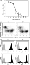
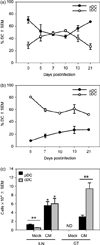
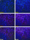
Dendritic cells (DCs) are central for the induction of T-cell responses needed for chlamydial eradication. Here, we report the activation of two DC subsets: a classical CD11b+ (cDC) and plasmacytoid (pDC) during genital infection with Chlamydia muridarum. Genital infection induced an influx of cDC and pDC into the genital tract and its draining lymph node (iliac lymph nodes, ILN) as well as colocalization with T cells in the ILN. Genital infection with C. muridarum also stimulated high levels of costimulatory molecules on cDC central for the activation of naïve T cells in vivo. In contrast, pDC expressed low levels of most costimulatory molecules in vivo and did not secrete cytokines associated with the production of T helper (Th)1 cells in vitro. However, pDC upregulated inducible costimulatory ligand expression and produced IL-6 and IL-10 in response to chlamydial exposure in vitro. Our findings show that these two DC subsets likely have different functions in vivo. cDCs are prepared for induction of antichlamydial T-cell responses, whereas pDCs have characteristics associated with the differentiation of non-Th1 cell subsets.
Figures


Similar articles
-
Plasmacytoid dendritic cells modulate nonprotective T-cell responses to genital infection by Chlamydia muridarum.FEMS Immunol Med Microbiol. 2010 Apr;58(3):397-404. doi: 10.1111/j.1574-695X.2010.00653.x. Epub 2010 Jan 19. FEMS Immunol Med Microbiol. 2010. PMID: 20180848 Free PMC article.
-
Prostaglandin E2 modulates dendritic cell function during chlamydial genital infection.Immunology. 2008 Feb;123(2):290-303. doi: 10.1111/j.1365-2567.2007.02642.x. Epub 2007 Aug 3. Immunology. 2008. PMID: 17680801 Free PMC article.
-
The infecting dose of Chlamydia muridarum modulates the innate immune response and ascending infection.Infect Immun. 2004 Nov;72(11):6330-40. doi: 10.1128/IAI.72.11.6330-6340.2004. Infect Immun. 2004. PMID: 15501762 Free PMC article.
-
Duration of untreated chlamydial genital infection and factors associated with clearance: review of animal studies.J Infect Dis. 2010 Jun 15;201 Suppl 2:S96-103. doi: 10.1086/652393. J Infect Dis. 2010. PMID: 20470047 Review.
-
Chlamydia Spreading from the Genital Tract to the Gastrointestinal Tract - A Two-Hit Hypothesis.Trends Microbiol. 2018 Jul;26(7):611-623. doi: 10.1016/j.tim.2017.12.002. Epub 2017 Dec 27. Trends Microbiol. 2018. PMID: 29289422 Free PMC article. Review.
Cited by
-
Plasmacytoid dendritic cells modulate nonprotective T-cell responses to genital infection by Chlamydia muridarum.FEMS Immunol Med Microbiol. 2010 Apr;58(3):397-404. doi: 10.1111/j.1574-695X.2010.00653.x. Epub 2010 Jan 19. FEMS Immunol Med Microbiol. 2010. PMID: 20180848 Free PMC article.
-
Interruption of CXCL13-CXCR5 axis increases upper genital tract pathology and activation of NKT cells following chlamydial genital infection.PLoS One. 2012;7(11):e47487. doi: 10.1371/journal.pone.0047487. Epub 2012 Nov 26. PLoS One. 2012. PMID: 23189125 Free PMC article.
-
Modulation of T helper 1 and T helper 2 immune balance in a murine stress model during Chlamydia muridarum genital infection.PLoS One. 2020 May 15;15(5):e0226539. doi: 10.1371/journal.pone.0226539. eCollection 2020. PLoS One. 2020. PMID: 32413046 Free PMC article.
-
Alternatively Activated Macrophages Are Host Cells for Chlamydia trachomatis and Reverse Anti-chlamydial Classically Activated Macrophages.Front Microbiol. 2019 May 7;10:919. doi: 10.3389/fmicb.2019.00919. eCollection 2019. Front Microbiol. 2019. PMID: 31134002 Free PMC article.
-
Significant roles played by IL-10 in Chlamydia infections.Inflammation. 2014 Jun;37(3):818-23. doi: 10.1007/s10753-013-9801-1. Inflammation. 2014. PMID: 24398613 Review.
References
-
- Ardavin C. Origin, precursors and differentiation of mouse dendritic cells. Nat Rev Immunol. 2003;3:582–590. - PubMed
-
- Asselin-Paturel C, Brizard G, Pin JJ, Briere F, Trinchieri G. Mouse strain differences in plasmacytoid dendritic cell frequency and function revealed by a novel monoclonal antibody. J Immunol. 2003;171:6466–6477. - PubMed
-
- Bilenki L, Wang S, Yang J, et al. Adoptive transfer of CD8alpha+ dendritic cells (DC) isolated from mice infected with Chlamydia muridarum are more potent in inducing protective immunity than CD8alpha- DC. J Immunol. 2006;177:7067–7075. - PubMed
Publication types
MeSH terms
Substances
Grants and funding
LinkOut - more resources
Full Text Sources
Medical
Research Materials
