. 2009 Jan 21:9:2.
doi: 10.1186/1471-2342-9-2.

Age and gender specific normal values of left ventricular mass, volume and function for gradient echo magnetic resonance imaging: a cross sectional study

Affiliations

Age and gender specific normal values of left ventricular mass, volume and function for gradient echo magnetic resonance imaging: a cross sectional study

Peter A Cain et al. BMC Med Imaging. .

Abstract

Background: Knowledge about age-specific normal values for left ventricular mass (LVM), end-diastolic volume (EDV), end-systolic volume (ESV), stroke volume (SV) and ejection fraction (EF) by cardiac magnetic resonance imaging (CMR) is of importance to differentiate between health and disease and to assess the severity of disease. The aims of the study were to determine age and gender specific normal reference values and to explore the normal physiological variation of these parameters from adolescence to late adulthood, in a cross sectional study.

Methods: Gradient echo CMR was performed at 1.5 T in 96 healthy volunteers (11-81 years, 50 male). Gender-specific analysis of parameters was undertaken in both absolute values and adjusted for body surface area (BSA).

Results: Age and gender specific normal ranges for LV volumes, mass and function are presented from the second through the eighth decade of life. LVM, ESV and EDV rose during adolescence and declined in adulthood. SV and EF decreased with age. Compared to adult females, adult males had higher BSA-adjusted values of EDV (p = 0.006) and ESV (p < 0.001), similar SV (p = 0.51) and lower EF (p = 0.014). No gender differences were seen in the youngest, 11-15 year, age range.

Conclusion: LV volumes, mass and function vary over a broad age range in healthy individuals. LV volumes and mass both rise in adolescence and decline with age. EF showed a rapid decline in adolescence compared to changes throughout adulthood. These findings demonstrate the need for age and gender specific normal ranges for clinical use.


Figures

Figure 1
Figure 1
Delineation of the left ventricular borders in the short axis plane in end-diastole and end-systole. Both the endocardial and epicardial borders were outlined manually for both ventricular volume and mass measurement (solid lines). Papillary muscle measurements were only included for mass measurements (dashed lines).
Figure 2
Figure 2
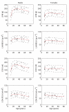
The normal age variation in left ventricular parameters in males (left) and females (right). Solid lines represent rational polynomial curve fit and dashed lines the 95% prediction intervals of this fit. Reference values are listed in Table 2. LVM = left ventricular mass, BSA = body surface area, EDV = end-diastolic volume.
Figure 3
Figure 3
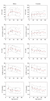
The normal age variation in left ventricular parameters in males (left) and females (right). Solid lines represent rational polynomial curve fit and dashed lines the 95% prediction intervals of this fit. Reference values are listed in Table 2. BSA = body surface area, ESV = end-systolic volume, SV = stroke volume, EF = ejection fraction.

References

    1. Pennell DJ, Sechtem UP, Higgins CB, Manning WJ, Pohost GM, Rademakers FE, van Rossum AC, Shaw LJ, Yucel EK. Clinical indications for cardiovascular magnetic resonance (CMR): Consensus Panel report. J Cardiovasc Magn Reson. 2004;6(4):727–765. doi: 10.1081/JCMR-200038581. - DOI - PubMed
    1. Hees PS, Fleg JL, Lakatta EG, Shapiro EP. Left ventricular remodeling with age in normal men versus women: novel insights using three-dimensional magnetic resonance imaging. Am J Cardiol. 2002;90(11):1231–1236. doi: 10.1016/S0002-9149(02)02840-0. - DOI - PubMed
    1. Lorenz CH, Walker ES, Morgan VL, Klein SS, Graham TP Jr. Normal human right and left ventricular mass, systolic function, and gender differences by cine magnetic resonance imaging. J Cardiovasc Magn Reson. 1999;1(1):7–21. doi: 10.3109/10976649909080829. - DOI - PubMed
    1. Marcus JT, DeWaal LK, Gotte MJ, Geest RJ van der, Heethaar RM, Van Rossum AC. MRI-derived left ventricular function parameters and mass in healthy young adults: relation with gender and body size. Int J Card Imaging. 1999;15(5):411–419. doi: 10.1023/A:1006268405585. - DOI - PubMed
    1. Sandstede J, Lipke C, Beer M, Hofmann S, Pabst T, Kenn W, Neubauer S, Hahn D. Age- and gender-specific differences in left and right ventricular cardiac function and mass determined by cine magnetic resonance imaging. Eur Radiol. 2000;10(3):438–442. doi: 10.1007/s003300050072. - DOI - PubMed

Publication types

MeSH terms

LinkOut - more resources