. 2008 Sep;10(9):1076-82.
doi: 10.1038/ncb1767.

Plk1-dependent phosphorylation of FoxM1 regulates a transcriptional programme required for mitotic progression

Affiliations

Plk1-dependent phosphorylation of FoxM1 regulates a transcriptional programme required for mitotic progression

Zheng Fu et al. Nat Cell Biol. 2008 Sep.

Abstract

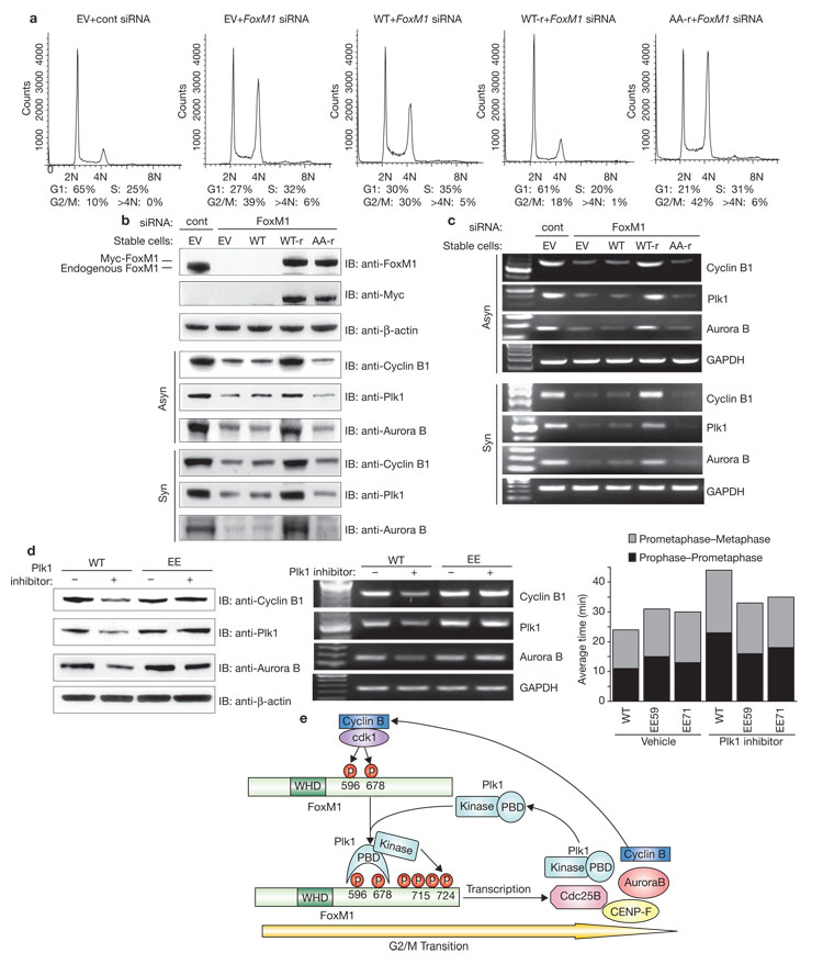
Proper control of entry into and progression through mitosis is essential for normal cell proliferation and the maintenance of genome stability. The mammalian mitotic kinase Polo-like kinase 1 (Plk1) is involved in multiple stages of mitosis5. Here we report that Forkhead Box M1 (FoxM1), a substrate of Plk1, controls a transcriptional programme that mediates Plk1-dependent regulation of cell-cycle progression. The carboxy-terminal domain of FoxM1 binds Plk1, and phosphorylation of two key residues in this domain by Cdk1 is essential for Plk1-FoxM1 interaction. Formation of the Plk1-FoxM1 complex allows for direct phosphorylation of FoxM1 by Plk1 at G2/M and the subsequent activation of FoxM1 activity, which is required for expression of key mitotic regulators, including Plk1 itself. Thus, Plk1-dependent regulation of FoxM1 activity provides a positive-feedback loop ensuring tight regulation of transcriptional networks essential for orderly mitotic progression.


Figures

Figure 1
Figure 1
Plk1 interacts with FoxM1 in vitro and in vivo. (a) In vitro translated FoxM1 was used in a pulldown assay with either GST or GST–Plk1 immobilized on agarose beads. (b) 293T cells were transfected with constructs encoding Myc-tagged FoxM1 and co-immunoprecipitation assays were performed. (c) Cell extracts were prepared from asynchronous (Asyn) or mitotic (M) HeLa cells and subjected to immunoprecipitation using anti-FoxM1 or anti-Plk1 antibodies. Immunoblotting was carried out using anti-Plk1 or anti-FoxM1 antibodies. (d) 293T cells were transfected with plasmids encoding Myc-tagged wild-type (FoxM1WT) or FoxM1 mutants (FoxM1T595A, FoxM1S678A or FoxM1TSAA). Cell lysates were subjected to pulldown assays using beads coated with GST–Plk1 PBD wild-type (WT) or a mutant (MUT) deficient in phosphopeptide binding. Beads coated with GST alone were used as a negative control. Full scans of blots in a–d are shown in Supplementary Information, Fig. S5.
Figure 2
Figure 2
Plk1 phosphorylates FoxM1 in vitro and in vivo. (a) Bacterially expressed FoxM1 was subjected to in vitro kinase assays. Kinases used were constitutively active human Plk1 (TD) or a kinase-defective (KD) mutant of Plk1 purified from insect cells. Loading controls are shown in the bottom panel (CBB, Coomassie blue staining). (b) FoxM1 transactivation domain mutants were used as substrates for in vitro kinase assays, similarly to that described in a. (c) Comparison of the extreme C-terminal sequence of FoxM1 orthologues. The two major phosphorylation sites, Ser 715 and Ser 724, are highlighted. (d) 293T cells were transfected with plasmids encoding Myc-tagged FoxM1WT or FoxM1S715A and FoxM1S724A mutants. Ectopically expressed FoxM1 proteins were immunoprecipitated with an anti-Myc antibody and immunoblotted with anti-p715, anti-p724 or anti-FoxM1 antibodies. (e) Endogenous FoxM1 was immunoprecipitated from cell lysates prepared from asynchronous (Asyn) or mitotic (M) HeLa cells. The phosphorylation status of FoxM1 was examined by western blotting using anti-p715 and anti-p724 antibodies. (f) HeLa cells were transfected with Plk1-specific siRNA (Plk1) or nonspecific control siRNA (Cont) against firefly luciferase. Nocodazole was added to arrest cells at mitosis 32 h after transfection. After a further 16 h of incubation, endogenous FoxM1 was immunoprecipitated and western blots were performed. Erk2 was included as a loading control. Full scans of blots in d–f are shown in Supplementary Information, Fig. S5.
Figure 3
Figure 3
Plk1 activates FoxM1 transcriptional activity. (a) U2OS cells were transiently transfected with the reporter plasmid 6×FoxM1 TATA–luciferase plasmid encoding wild-type FoxM1, and increasing amounts of plasmids encoding for either wild-type Plk1 (Plk1WT) or kinase-defective Plk1 (Plk1KD). (b) U2OS cells were transfected with the FoxM1 reporter plasmid and plasmids encoding for either wild-type FoxM1 (FoxM1WT) or FoxM1 mutants (FoxM1T596A, FoxM1S678A or FoxM1TSAA) together with plasmids encoding for WT or KD Plk1. (c) U2OS cells were transfected with the FoxM1 reporter plasmid and plasmids encoding for either Myc-tagged FoxM1WT, FoxM1 mutants (FoxM1AA, FoxM1EE), or a FoxM1 TAD-deletion mutant (FoxM11–616) together with Plk1 WT or KD expression vectors. Luciferase activities were measured 24 h after transfection. Luciferase levels were normalized to Renilla luciferase activity. The data are expressed as a percentage of wildtype FoxM1 activity (mean ± s.d. of three separate experiments in triplicate). Expression levels of exogenous wild-type and mutant FoxM1 were monitored by western blot analysis.
Figure 4
Figure 4
Plk1-dependent phosphorylation of FoxM1 is required for expression of the G2/M transcriptional programme and orderly mitotic progression in vivo. U2OS cells stably expressing empty vector (EV), or Myc-tagged FoxM1WT, siRNA-resistant wild-type FoxM1 (WT-r) or siRNA-resistant S715A;S724A mutant FoxM1 (AA-r) were transfected with FoxM1-specific siRNA (FoxM1) or nonspecific control siRNA (cont). (a) Cell-cycle distributions were determined by flow cytometry analysis 72 h after transfection. (b) Lysates were collected 72 h after transfection and analysed by immunoblotting (Asyn). Alternatively, cells were synchronized by nocodazole treatment for 16 h before collection (Syn). Blotting for β-actin was used as a loading control. (c) Total RNA was extracted 48 h after transfection and subjected to RT–PCR analysis. RT–PCR for GAPDH was used as an internal control. (d) U2OS cells stably expressing FoxM1 wildtype (WT) or phosphomimetic mutants (EE) were synchronized by thymidine block and released in the presence of either vehicle (DMSO) or ON01910 (250 nM) for 9 h. Cells were subjected to RT–PCR and immunoblotting analysis or monitored by time-lapse imaging. Two independent stable clones (EE59 and EE71) were used in time-lapse microscopy. Inhibition of Plk1 activity led to 91% of wild-type FoxM1-expressing cells arresting at prometaphase, which was partially rescued in phosphomimetic FoxM1-expressing cells (EE59, 67%, EE71, 68%). For cells that eventually progressed through mitosis, the duration of each stage of mitosis was recorded and average time was plotted in the bar chart. (e) Model for Plk1-mediated regulation of FoxM1 transcriptional activity at G2/M transition.

References

    1. Nigg EA. Mitotic kinases as regulators of cell division and its checkpoints. Nature Rev. Mol. Cell Biol. 2001;2:21–32. - PubMed
    1. Norbury C, Nurse P. Animal cell cycles and their control. Annu. Rev. Biochem. 1992;61:441–470. - PubMed
    1. Pines J, Rieder CL. Re-staging mitosis: a contemporary view of mitotic progression. Nature Cell Biol. 2001;3:E3–E6. - PubMed
    1. Murray AW. Recycling the cell cycle: cyclins revisited. Cell. 2004;116:221–234. - PubMed
    1. Barr FA, Sillje HH, Nigg EA. Polo-like kinases and the orchestration of cell division. Nature Rev. Mol. Cell Biol. 2004;5:429–440. - PubMed

Publication types