

doi: 10.1038/nn.2178.
Pubertal hormones modulate the addition of new cells to sexually dimorphic brain regions
Affiliations
- PMID: 19160494
- PMCID: PMC2772186
- DOI: 10.1038/nn.2178
Item in Clipboard
Pubertal hormones modulate the addition of new cells to sexually dimorphic brain regions
Nat Neurosci.
2008 Sep.
Abstract
New cells, including neurons, arise in several brain regions during puberty in rats. Sex differences in pubertal addition of cells coincide with adult sexual dimorphisms: for each region, the sex that gains more cells during puberty has a larger volume in adulthood. Removing gonadal hormones before puberty eliminates these sex differences, indicating that gonadal steroids direct the addition of new cells during puberty to maintain and accentuate sexual dimorphisms in the adult brain.
Figures
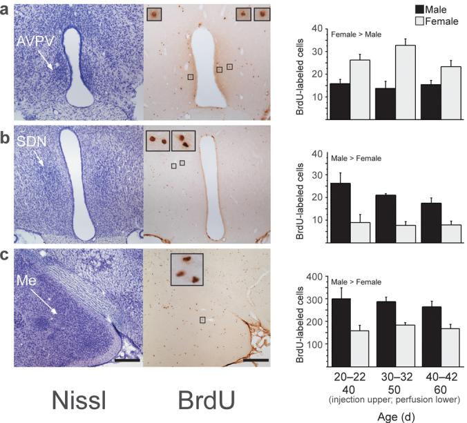
New cells are added during puberty to the AVPV (a), SDN (b), and Me (c) in male and female rats. Left photomicrographs are of thionin-stained sections and right photomicrographs are of BrdU-labeled cells in nearby sections from the same animal. Insets show BrdU-labeled cells framed in small boxes at 10x higher magnification. Subjects received a daily injection of 300 mg/kg BrdU on three consecutive days on either 20-22, 30-32, or 40-42 days of age (n=6-8/age and sex). BrdU is incorporated into DNA during the S phase of the cell cycle and can be later visualized to identify cells replicating at the time of BrdU administration. Brain tissue was collected 20 days after the first BrdU injection, on 40, 50, or 60 days of age, respectively. All protocols involving animals were approved by the Michigan State University Institutional Animal Care and Use Committee. Quantitative analyses of BrdU-labeled cells revealed that during puberty, significantly more cells were added to AVPV (a) in females than in males, while significantly more cells were added to SDN (b) and Me (c) in males than in females. Data are means ± SEM. Scale bars: 250 μm in lower magnification images.
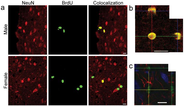
Many BrdU-labeled cells in the AVPV are mature neurons, not astrocytes. Many BrdU-labeled cells in Me are astrocytes; some are neurons. a: Confocal images of cells in male and female rat AVPV. Sections were processed for double-label BrdU (green) and the mature neuron marker NeuN (red); colocalization is yellow. b: Orthogonal views of confocal images verify colocalization of BrdU and NeuN in the female AVPV of a neuron that was born on P30-32. c: Confocal image of cells in rat Me. Section was processed for triple-label BrdU (green), NeuN (blue), and the astrocytic glial marker GFAP (red). Orthogonal views verify colocalization of BrdU, which is in the nucleus, and GFAP, which is in astrocytic processes. Scale bars: 10 μm.
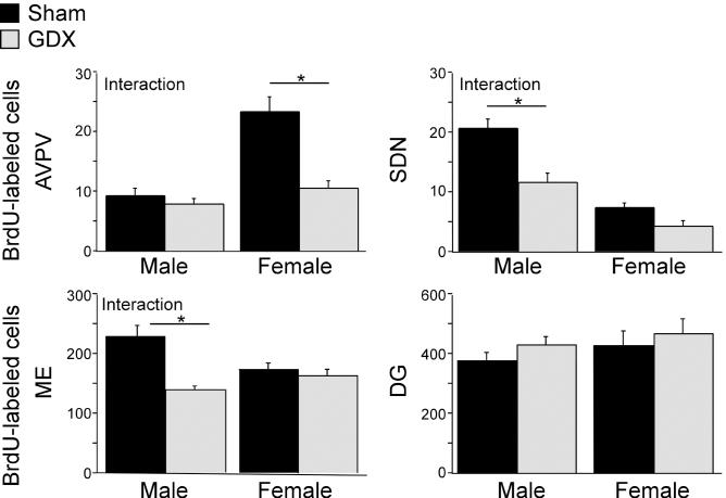
The effect of prepubertal gonadectomy (GDX) on the number of BrdU-labeled cells depends on sex and brain region. Male and female rats were gonadectomized or sham gonadectomized at 20 days of age (n=8/sex and treatment). A daily injection of BrdU was given on 30-32 days of age and brain tissue was collected at 50 days of age. Prepubertal GDX significantly decreased the number of BrdU-labeled cells in female but not male AVPV (interaction between sex and treatment). Prepubertal GDX decreased the number of BrdU-labeled cells in male but not female SDN and Me (interaction between sex and treatment). Prepubertal GDX did not affect BrdU-labeled cells in the dentate gyrus (DG) of either males or females. Data are presented as means ± SEM. Asterisks indicate p<0.05 (post hoc Fisher test between groups).
References
Publication types
MeSH terms
Substances
Grants and funding
LinkOut - more resources
Full Text Sources
Other Literature Sources
Medical
