T(2) relaxometry of normal pediatric brain development
- PMID: 19161173
- PMCID: PMC2767196
- DOI: 10.1002/jmri.21646
T(2) relaxometry of normal pediatric brain development
Abstract
Purpose: To establish normal age-related changes in the magnetic resonance (MR) T(2) relaxation time constants of brain using data collected as part of the National Institutes of Health (NIH) MRI Study of Normal Brain Development.
Materials and methods: This multicenter study of normal brain and behavior development provides both longitudinal and cross-sectional data, and has enabled us to investigate T(2) evolution in several brain regions in healthy children within the age range of birth through 4 years 5 months. Due to the multicenter nature of the study and the extended period of data collection, periodically scanned inanimate and human phantoms were used to assess intra- and intersite variability.
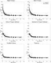
Results: The main finding of this work, based on over 340 scans, is the identification and parameterization of the monoexponential evolution of T(2) from birth through 4 years 5 months of age in various brain structures.
Conclusion: The exponentially decaying T(2) behavior is believed to reflect the rapid changes in water content as well as myelination during brain development. The data will become publicly available as part of a normative pediatric MRI and clinical/behavioral database, thereby providing a basis for comparison in studies assessing normal brain development, and studies of deviations due to various neurological, neuropsychiatric, and developmental disorders.
Figures


Comment in
-
T2 relaxometry of maturing brains.J Magn Reson Imaging. 2009 Oct;30(4):911; author reply 912. doi: 10.1002/jmri.21927. J Magn Reson Imaging. 2009. PMID: 19787741 No abstract available.
References
-
- Paus T, Collins DL, Evans AC, Leonard G, Pike B, Zijdenbos A. Maturation of white matter in the human birth: A review of magnetic resonance studies. Brain Research Bulletin. 2001;54:255–266. - PubMed
-
- Takeda K, Nomura Y, Sakuma H, Tagami T, Okuda Y, Nakagawa T. MR assessment of normal brain development in neonates and infants: comparative study of T1- and diffusion-weighted images. J Comput Assist Tomogr. 1997;21:1–7. - PubMed
-
- Van der Knaap MS, Valk J. MR imaging of the various stages of normal myelination during the first year of life. Neuroradiology. 1990;31:459–470. - PubMed
Publication types
MeSH terms
Grants and funding
- N01 NS092315/NS/NINDS NIH HHS/United States
- N01-HD02-3343/HD/NICHD NIH HHS/United States
- N01-NS-9-2319/NS/NINDS NIH HHS/United States
- N01-NS-9-2315/NS/NINDS NIH HHS/United States
- N01 MH090002/MH/NIMH NIH HHS/United States
- N01 NS092317/NS/NINDS NIH HHS/United States
- N01 NS092319/NS/NINDS NIH HHS/United States
- N01 NS092316/NS/NINDS NIH HHS/United States
- N01-NS-9-2316/NS/NINDS NIH HHS/United States
- N01-NS-9-2320/NS/NINDS NIH HHS/United States
- N01-MH9-0002/MH/NIMH NIH HHS/United States
- N01-NS-9-2314/NS/NINDS NIH HHS/United States
- N01 NS092320/NS/NINDS NIH HHS/United States
- N01 HD023343/HD/NICHD NIH HHS/United States
- N01-NS-9-2317/NS/NINDS NIH HHS/United States
- N01 NS092314/NS/NINDS NIH HHS/United States
LinkOut - more resources
Full Text Sources
Other Literature Sources
Medical

