. 2009 Feb;238(2):431-42.
doi: 10.1002/dvdy.21854.

Ligand-specific function of transforming growth factor beta in epithelial-mesenchymal transition in heart development

Affiliations

Ligand-specific function of transforming growth factor beta in epithelial-mesenchymal transition in heart development

Mohamad Azhar et al. Dev Dyn. 2009 Feb.

Abstract

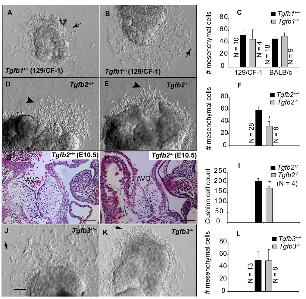
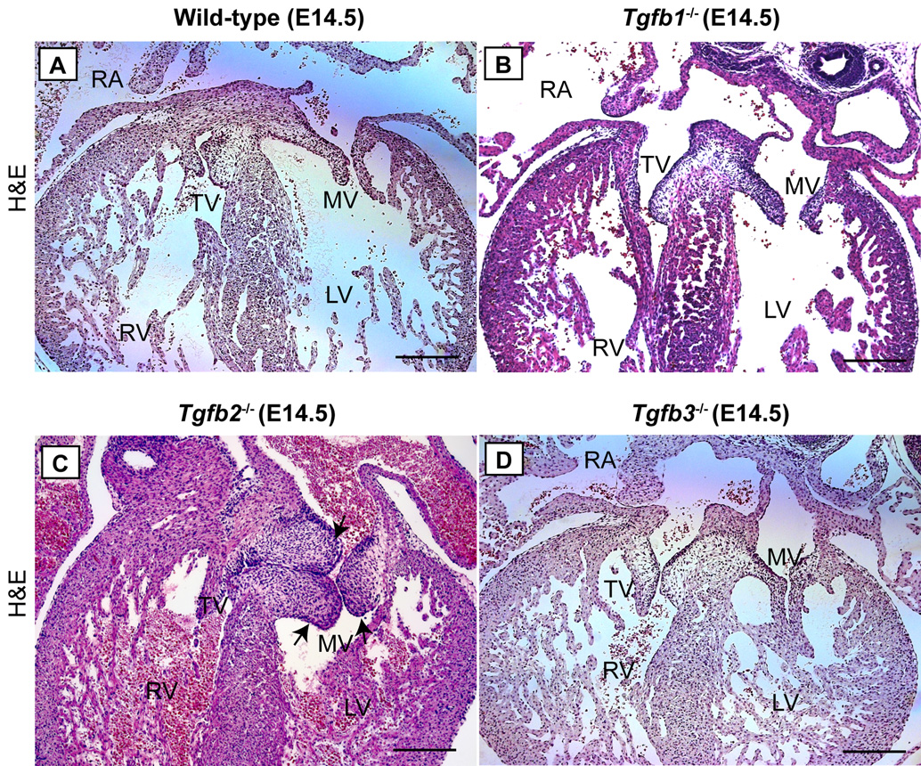
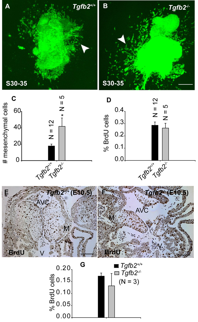
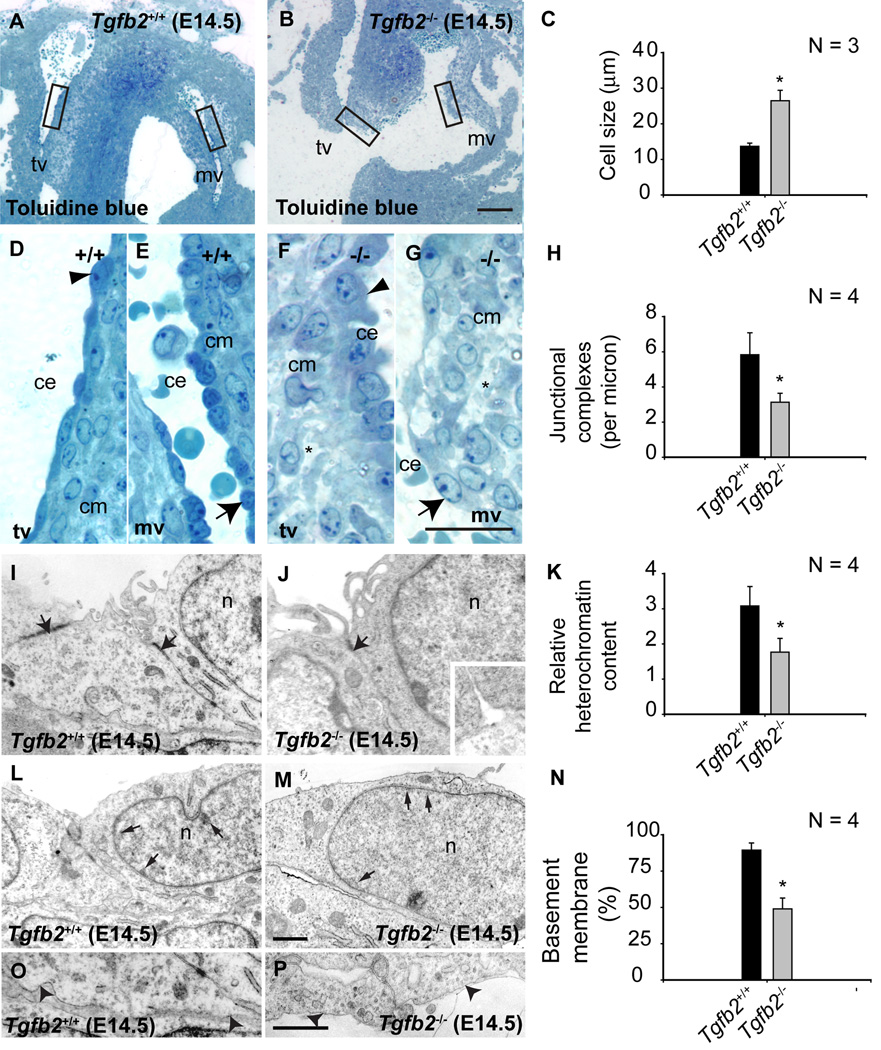
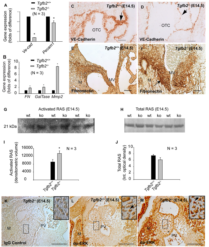
The ligand specificity of transforming growth factor beta (TGFbeta) in vivo in mouse cardiac cushion epithelial-to-mesenchymal transition (EMT) is poorly understood. To elucidate the function of TGFbeta in cushion EMT, we analyzed Tgfb1(-/-), Tgfb2(-/-), and Tgfb3(-/-) mice between embryonic day (E) 9.5 and E14.5 using both in vitro and in vivo approaches. Atrioventricular (AV) canal collagen gel assays at E9.5 indicated normal EMT in both Tgfb1(-/-) and Tgfb3(-/-) mice. However, analysis of Tgfb2(-/-) AV explants at E9.5 and E10.5 indicated that EMT, but not cushion cell proliferation, was initially delayed but later remained persistent. This was concordant with the observation that Tgfb2(-/-) embryos, and not Tgfb1(-/-) or Tgfb3(-/-) embryos, develop enlarged cushions at E14.5 with elevated levels of well-validated indicators of EMT. Collectively, these data indicate that TGFbeta2, and not TGFbeta1 or TGFbeta3, mediates cardiac cushion EMT by promoting both the initiation and cessation of EMT.


Figures

Fig.1
Fig.1. Effect of gene targeted disruption of Tgfb1, Tgfb2 and Tgfb3 on cushion EMT at an early stage
Representative images of in vitro AV canal collagen gel assays (A,B,D,E,J,K) of Tgfb1+/+ (A) and Tgfb1−/− (B), Tgfb2+/+ (D) and Tgfb2−/− (E), and Tgfb3+/+ (J) and Tgfb3−/− (K) embryos at S21–29 (E9.5). Images were taken after 24 hr of explant culturing. Note that the number of mesenchymal cells present in the collagen gels cannot be determined with precision from the micrograph as many of the mesenchymal cells in our explant cultures were found beneath endothelial cells, and therefore they were not captured in the micrographs. C,F,L: Quantification of the number of mesenchymal cells (mean±s.e.m) below the gel surface (e.g., arrows (A,B,J,K) or arrowheads (A,B)). The number of mesenchymal cells is not significantly changed in Tgfb1−/− mice (C) (P=0.6640 for 129/CF-1, P= 0.4573 for BALB/c) or Tgfb3−/− mice (L) (P= 0.9737), but it is significantly reduced in Tgfb2−/− mice (F) (*P=0.0203). G-I: H-E stained images of showing AV cushions of E10.5 wild-type (G) and Tgfb2−/− mice (H) and the relative total cushion cell count in these embryos (I). The total cushion count is significantly reduced in Tgfb2−/− cushions (*P= 0.0201). Images are representative of 4 wild-type/mutant embryo pairs. Scale bar in (J ) applies to (A-B, D-E, J-K) = 100 µm; (G,H) = 100µm. AVC, AV cushions.
Fig.2
Fig.2. Cushion morphology in Tgfb1−/−, Tgfb2−/− and Tgfb3−/− mice at E14.5
A-D: Representative of serially sectioned H-E stained images of wild-type (A), Tgfb1−/− (B), Tgfb2−/− (C) and Tgfb3−/− (D). Only Tgfb2−/− mice develop dysplastic or enlarged cushions (C, arrows) (see Results section for a quantification of total cushion cells). Notably, this mutant heart also has overriding tricuspid valves. Scale bar: 200 µm (A–D). TV, tricuspid valves; MV, mitral valves; RA, right atrium; RV, right ventricle; LV, left ventricle.
Fig.3
Fig.3. Effect of gene targeted disruption of Tgfb2 on cushion EMT and cell proliferation after the rudimentary cushions are formed at E10.5
A-B: Representative confocal images of alexa-fluor488-phalloidin stained S30–35 (E10.5) AV canal explants of Tgfb2+/+ (A) and Tgfb2−/− (B) embryos after 60 hr of culturing. The AV explants in which the epicardial cell layer is removed or destroyed at the time of harvesting are cultured for 12 hr to let the pre-existing cushion mesenchymal cells migrate out of the AV explants and then transferred to fresh collagen gels for an additional 48 hr. (C) The number of mesenchymal cells (mean±s.e.m) below the gel surface (arrowhead, a-b). Notably, as the endocardium and epicardium are the only possible sources of mesenchyme in this experiment, differences between the wild type and Tgfb2−/− mice reflect the unimpeded endothelial source and, perhaps, a few cells that may have escaped the epicardial disruption procedure (see Experimental Procedures for details). (D) % of BrdU-incorporated mesenchymal cells (mean±s.e.m) in vitro on collagen gels. The number of mesenchymal cells is significantly increased in Tgfb2−/− mice (C) (*P=0.0032) but there is no difference in % of BrdU cells between wild-type and mutants (D). e-g: Anti-BrdU-stained sections of E10.5 AV cushions of wild-type (E) and Tgfb2−/− (F) mice and the % of BrdU-incorporating cushion cells (G) (brown color staining in E-F). Cell proliferation is comparable between wild-type and mutant cushions at E10.5 (P=0.9416). Images (E,F) are representative of three wild-type/mutant embryo pairs. Scale bar: 100 µm (A,B,E,F).
Fig.4
Fig.4. Cellular and ultrastructural changes in cushion endothelial cells in Tgfb2 −/− mice at E14.5
A-G: Toluidine blue staining of wild-type (A) and Tgfb2−/− (B) AV cushions. Boxes in (A) and (B) are magnified in (D,E) and (F,G), respectively. Asterisks (F,G) indicate the abnormal organization and composition of cardiac jelly in mutants. Cushion endothelial cells are enlarged in mutants (arrowheads (D,F) and arrows (E,G)). C: Morphometric comparison showing significantly increased cushion endothelial cell size (mean±s.e.m) in Tgfb2−/− embryos (*P= 0.0138). I-Q: Ultrastructural and morphometric comparison of wild-type and Tgfb2−/− mice by transmission electron microscopy. Electron micrographs of representative E14.5 Tgfb2+/+ (I,L,P) and Tgfb2−/− (J,M,Q) mice, illustrating decreased endothelial junctional complexes (arrows, I,J), heterochromatin organization (arrows, L,M), and basement membrane (arrowheads, P,Q). Morphometric comparison (mean±s.e.m) of junctional complexes (H), relative heterochromatin content (K) and % of basement membrane (O) confirms these findings. *P=0.0320 (junctional complexes), 0.0306 (heterochromatin content), 8.8927e-5 (basement membrane). Images (A,B,D-G) and (I,J,L,M,P,Q) are representative of four wild-type/mutant embryo pairs. Scale: 100 µm (a,b), 25 µm (D,E,F,G), 1 µm (scale in Q applies to I,J,L,M,P). mv, mitral valves; tv, tricuspid valves; ce, cushion endothelium; cm, cushion mesenchyme; n, nucleus.
Fig.5
Fig.5. Cushion EMT marker expression in Tgfb2−/− mice at E14.5
A-B: Q-RT-PCR (mean±s.e.m.) showing significantly decreased VE-Cad (*P=7.0000e-10) and Pecam1 (*P=0.0392) (A), and increased Fibronectin (*P = 0.0016), GalTase (*P=2.6790e-4) and Mmp2 (*P=6.4998e-6) (B) in AV explant tissue of Tgfb2−/− mice. Each wild-type value is normalized to 1.0. C-D: Anti-VE-Cadherin staining of wild-type (C) and Tgfb2−/− (D) mice shows that VE-Cadherin is reduced in mutants (arrows, C-D). E-F: Anti-FN staining of wild-type (E) and Tgfb2−/− (F) mice confirms the increased FN in AV cushions of Tgfb2−/− mice (arrow, F). Note that Tgfb2−/− cushions in (F) are naturally enlarged. G,H: Representative western blots showing increased activated RAS (G) and unaltered total RAS (H) in Tgfb2−/− hearts. The blots are a representative of at least 3 wild-type/mutant embryo pairs. I,J: Densitometric quantification (mean±s.e.m) confirms increased activated RAS in mutants (I) (*P=0.0394) without any significant change in the total RAS (J) (P=0.1710). K-M: Tgfb2−/− cushions stained with IgG (K) and anti-dp-ERK (L) are compared with anti-dp-ERK stained cushions of Tgfb2+/+ mice (M). Areas under the box in (K–M) are magnified (insets). Tgfb2−/− cushions have increased dp-ERK (arrows, L-M). Images (C-D, E-F, K-M) are representative of at least three wild-type/mutant embryo pairs. Scale bar: 50 µm (C-D, E-F, K-M). OTC, outflow tract cushion; MV, mitral valves; M, myocardium; PV, pulmonary valves.

References

    1. Abdelwahid E, Pelliniemi LJ, Jokinen E. Cell death and differentiation in the development of the endocardial cushion of the embryonic heart. Microsc Res Tech. 2002;58:395–403. - PubMed
    1. Ahmed S, Nawshad A. Complexity in interpretation of embryonic epithelial-mesenchymal transition in response to transforming growth factor-beta signaling. Cells Tissues Organs. 2007;185:131–145. - PMC - PubMed
    1. Armstrong EJ, Bischoff J. Heart valve development: endothelial cell signaling and differentiation. Circ Res. 2004;95:459–470. - PMC - PubMed
    1. Azhar M, Schultz JE, Grupp I, Dorn GW, Meneton P, Molin DG, Gittenberger-de Groot AC, Doetschman T. Transforming growth factor beta in cardiovascular development and function. Cytokine Growth Factor Rev. 2003;14:391–407. - PMC - PubMed
    1. Barnett JV, Desgrosellier JS. Early events in valvulogenesis: a signaling perspective. Birth Defects Res C Embryo Today. 2003;69:58–72. - PubMed

Publication types

MeSH terms

Substances