. 2009 Apr 14;48(14):3230-8.
doi: 10.1021/bi802163p.

Dissociation of the subunits of the calcium-independent receptor of alpha-latrotoxin as a result of two-step proteolysis

Affiliations

Dissociation of the subunits of the calcium-independent receptor of alpha-latrotoxin as a result of two-step proteolysis

Valery Krasnoperov et al. Biochemistry. .

Abstract

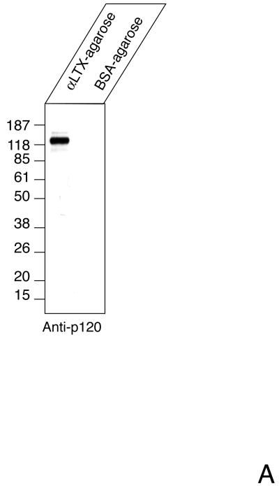
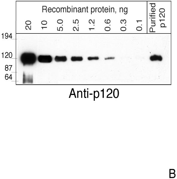
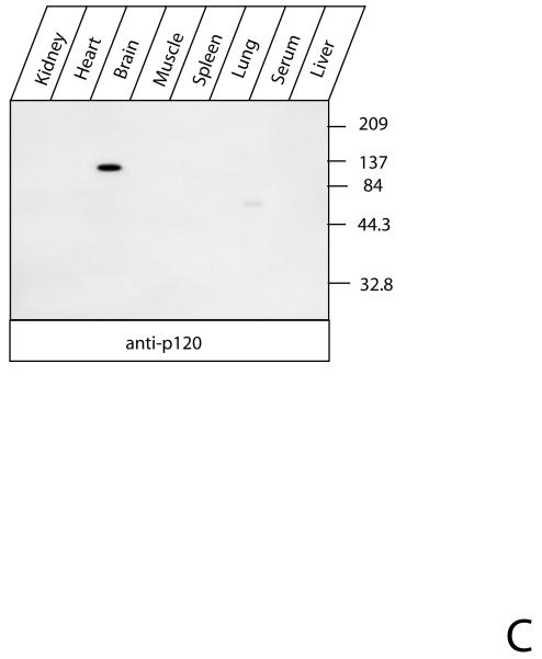
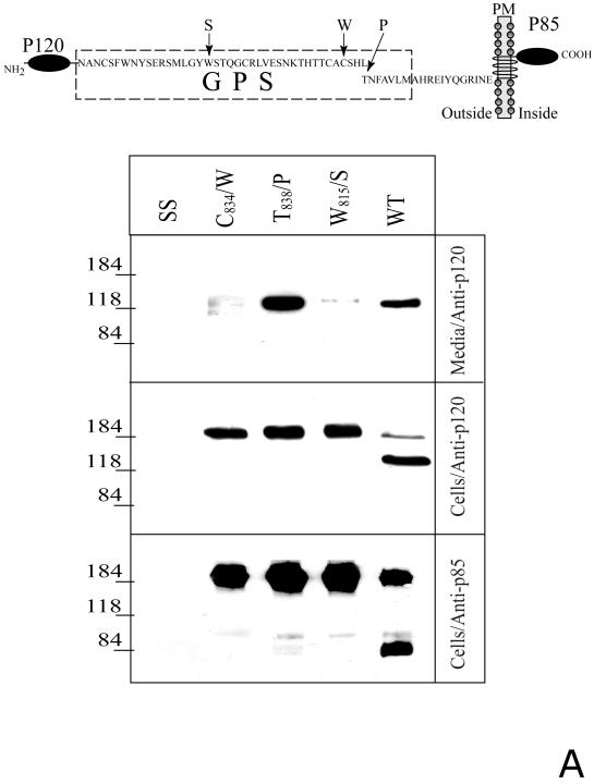
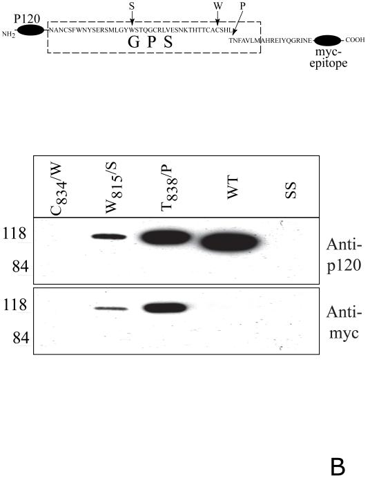
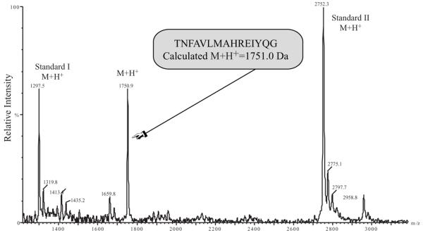
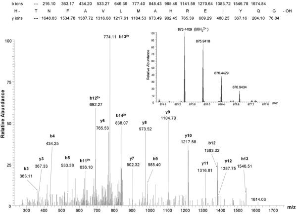
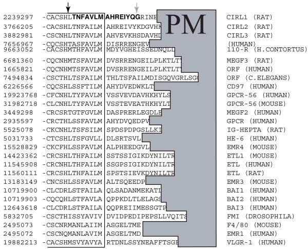
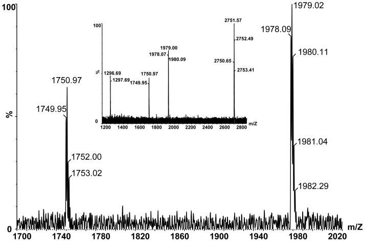
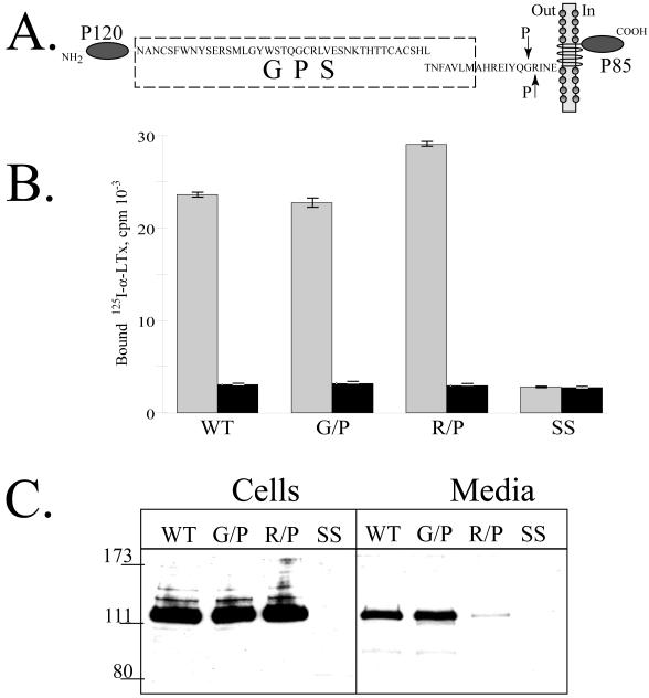
CIRL (the calcium-independent receptor of alpha-latrotoxin), a neuronal cell surface receptor implicated in the regulation of exocytosis, is a member of the GPS family of chimeric cell adhesion/G protein-coupled receptors. The predominant form of CIRL is a membrane-bound complex of two subunits, p120 and p85. Extracellularly oriented p120 contains hydrophilic cell adhesion domains, whereas p85 is a heptahelical membrane protein. Both subunits are encoded by the same gene and represent products of intracellular proteolytic processing of the CIRL precursor. In this study, we demonstrate that a soluble form of CIRL also exists in vitro and in vivo. It results from the further cleavage of CIRL by a second protease. The site of the second cleavage is located in the short N-terminal extracellular tail of p85, between the GPS domain and the first transmembrane segment of CIRL. Thus, the soluble form of CIRL represents a complex of p120 noncovalently bound to a 15 amino acid residue N-terminal peptide fragment of p85. We have previously shown that mutations of CIRL in the GPS domain inhibit intracellular proteolytic processing and also result in the absence of the receptors from the cell surface. Our current data suggest that although CIRL trafficking to the cell membrane is impaired by mutations in the GPS region, it is not blocked completely. However, at the cell surface, the noncleaved mutants are preferentially targeted by the second protease that sheds the extracellular subunit. Therefore, the two-step proteolytic processing may represent a regulatory mechanism that controls cell surface expression of membrane-bound and soluble forms of CIRL.


Figures

Fig. 1
Fig. 1. The soluble form of CIRL in brain
A. p120 in the soluble fraction of brain protein extract. Rat brains were extracted, precipitated with either α-latrotoxin-agarose or BSA-agarose and analyzed by Western blotting with anti-p120 antibody as described in Experimental Procedures. B. Semi-quantitative assay of soluble p120. One ml of rat brain extract equivalent to 8 mg of membrane protein was precipitated with 25 μl of α-latrotoxin-agarose for 15 hr at 4°C and analyzed by Western blotting with anti-p120 antibody. The indicated amounts of purified recombinant p120 were used as standards. C. Tissue distribution of soluble CIRL. Rat tissues were extracted and analyzed for the presence of p120 as described in the Experimental Procedures. The position of molecular mass protein standards (Gibco/Invitrogen) and their molecular mass in kDa are shown.
Fig. 2
Fig. 2. Proteolytic processing of the full-length and soluble deletion constructs of CIRL mutated in the GPS domain
A. Soluble forms of CIRL GPS mutants. COS cells were transfected with either wild-type CIRL or its GPS mutants with single residue substitution within the GPS domain (C834/W, W815/S, and T838/P), schematically described at the top panel, PM - plasma membrane. The cells were harvested and analyzed by Western blotting with anti-p120 or anti-p85 antibody. The conditioned media were precipitated with α-latrotoxin-agarose followed by Western blotting with anti-p120 antibody. Salmon sperm DNA transfected cells (SS) were used as control. B. Secretion of soluble GPS mutants of the CIRL ectodomain. COS cells were transfected with the plasmids encoding either the wild type CIRL ectodomain or three single-residue mutant constructs (C834/W, W815/S, and T838/P), schematically described at the top panel. The conditioned media were precipitated with α-latrotoxin-agarose followed by Western blotting with either anti-p120 or anti-myc antibody. Note an increase in the apparent size of p120 in the non-cleaved mutants W815/S, and T838/P due to the 3.8 kDa myc-tag addition. The pictures shown are representative of five independent transfection, precipitation and blotting experiments that produced essentially similar results.
Fig. 3
Fig. 3. Secondary proteolysis of CIRL-1
MALDI-TOF spectrum of the peptide product of the dual cleavage of CIRL by intracellular and extracellular proteases. The medium of CIRL-transfected COS cells (100 ml) was concentrated and precipitated with α-latrotoxin-agarose. The adsorbed material was eluted and analyzed by MALDI-TOF mass spectrometry. Internal mass standards were angiotensin I (average M+H+ 1297.5) and a synthetic peptide (average M+H+ 2752.3).
Fig. 4
Fig. 4. LTQ-Orbitrap LC-MS/MS spectrum of 7% of the eluate from the LTX affinity column using the soluble fraction of brain homogenate as starting material
Observed b and y ions from the peptide of sequence TNFAVLMAHREIYQG are labeled in the MS/MS spectrum. Inset shows a portion of the Orbitrap MS survey scan containing the doubly charged precursor ion of the sequenced peptide, which has a calculated m/z of 875.4407 (mass error 0.2 ppm).
Fig. 5
Fig. 5. Multiple alignment of GPS receptors in the region of the sites of intracellular and extracellular proteolysis
GenBank™ protein accession numbers are shown in the left column. The intracellular cleavage site identified in CIRL-1 and several other GPS receptors is shown by a black arrow. The site of the extracellular cleavage of CIRL-1 only is marked by a gray arrow. PM, plasma membrane, denotes the region of hydrophobic residues of the first transmembrane segments of the aligned receptors.
Fig. 6
Fig. 6. Secondary proteolysis of CIRL-2
Reflectron mode MALDI-TOF MS spectra of CIRL-2 cleavage peptide (calculated m/z of [M+H]+ ion = 1978.02). Large figure shows expanded view of an externally calibrated spectrum, inset shows a spectrum acquired with angiotensin I (calculated m/z of [M+H]+ ion = 1296.69) and a synthetic peptide (calculated m/z of [M+H]+ ion = 2750.65) as internal calibrants.
Fig. 7
Fig. 7. Cell surface expression of CIRL mutated at the second cleavage site
A. Schematic description of the CIRL constructs with single residue mutations (G852/P and R853/P, indicated by arrows) at the second cleavage site. B. Cell surface expression of the second cleavage site CIRL mutants. Intact COS cells transfected with either wild-type CIRL or its mutants (G852/P and R853/P) were assayed for binding of 125I-α-latrotoxin either in the presence (black bars) or absence of excess non-labeled α-latrotoxin. Measurements were performed in triplicates. C. Secretion of the soluble forms of CIRL mutants. One ml of conditioned media of the same cells as in B was precipitated with 10 μl of α-latrotoxin-agarose and the eluates were analyzed by Western blotting with anti-p120 antibody. SS, salmon sperm DNA transfected cells. The picture shown is representative of three independent transfection, precipitation and blotting experiments that produced essentially similar results.

References

    1. Hayflick JS. A family of heptahelical receptors with adhesion-like domains: a marriage between two super families. J Recept Signal Transduct Res. 2000;20:119–131. - PubMed
    1. Stacey M, Lin HH, Gordon S, McKnight AJ. LNB-TM7, a group of seven-transmembrane proteins related to family-B G-protein-coupled receptors. Trends in biochemical sciences. 2000;25:284–289. - PubMed
    1. Yona S, Lin HH, Siu WO, Gordon S, Stacey M. Adhesion-GPCRs: emerging roles for novel receptors. Trends in biochemical sciences. 2008;33:491–500. - PubMed
    1. Kwakkenbos MJ, Kop EN, Stacey M, Matmati M, Gordon S, Lin HH, Hamann J. The EGF-TM7 family: a postgenomic view. Immunogenetics. 2004;55:655–666. - PubMed
    1. Usui T, Shima Y, Shimada Y, Hirano S, Burgess RW, Schwarz TL, Takeichi M, Uemura T. Flamingo, a seven-pass transmembrane cadherin, regulates planar cell polarity under the control of Frizzled. Cell. 1999;98:585–595. - PubMed

Publication types

LinkOut - more resources