Review
. 2009 Jan-Feb;85(1):21-32.
doi: 10.1111/j.1751-1097.2008.00507.x.

Gold nanorods as contrast agents for biological imaging: optical properties, surface conjugation and photothermal effects

Affiliations
Review

Gold nanorods as contrast agents for biological imaging: optical properties, surface conjugation and photothermal effects

Ling Tong et al. Photochem Photobiol. 2009 Jan-Feb.

Abstract

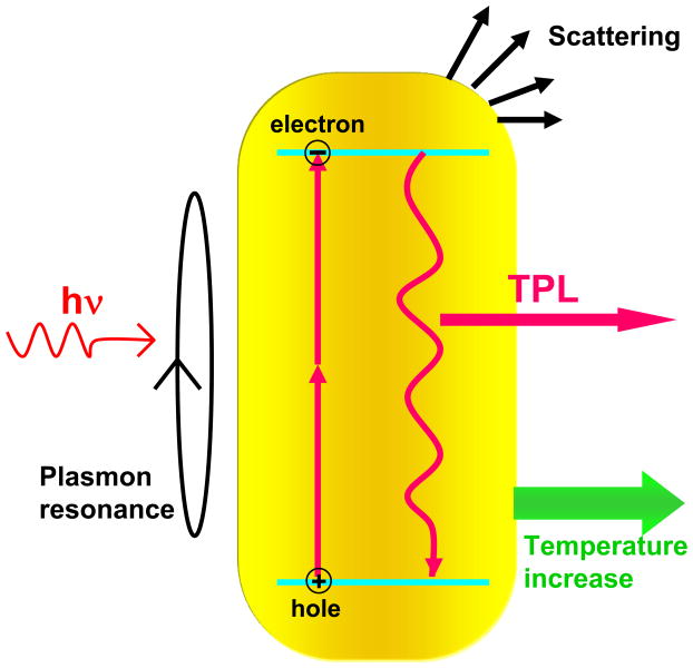
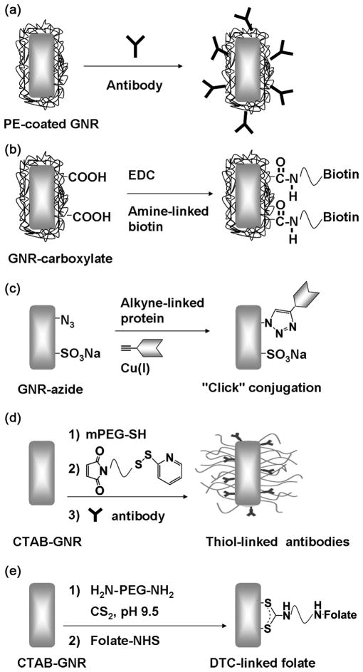
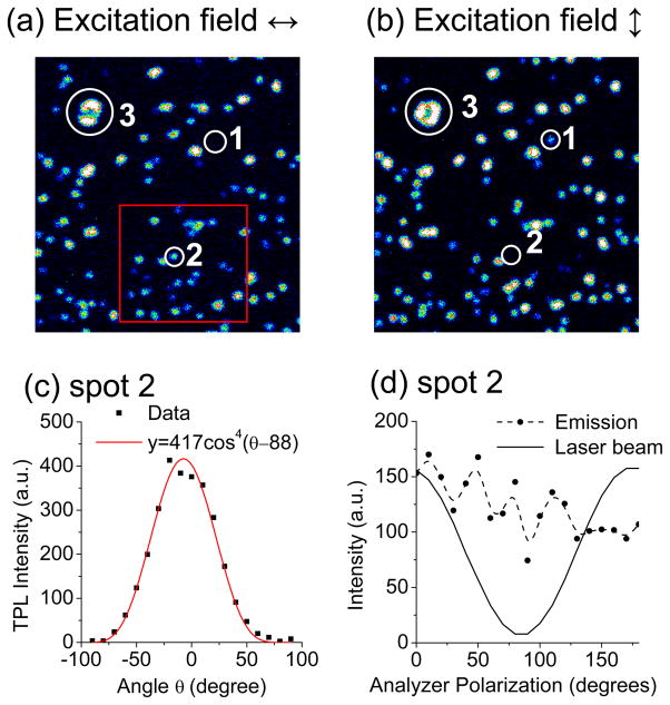
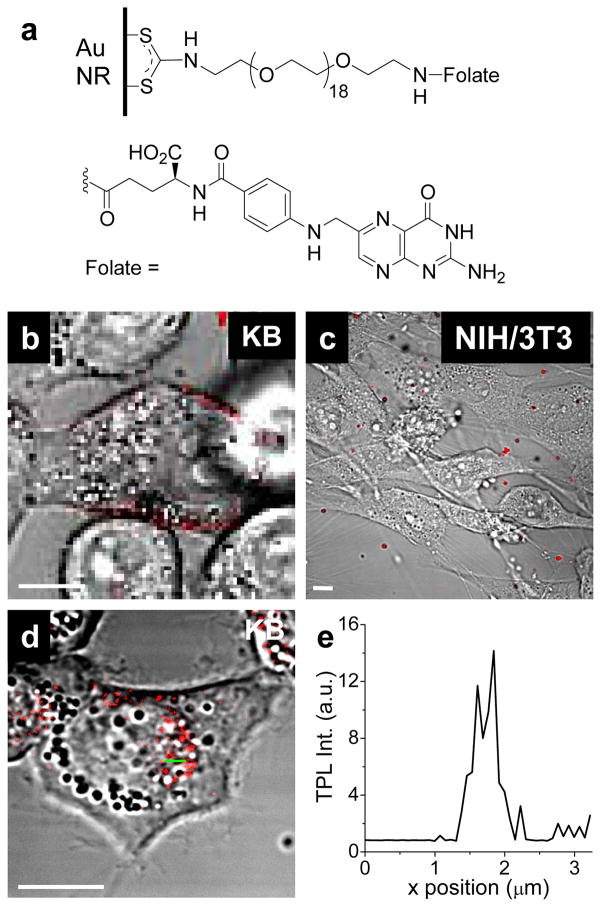
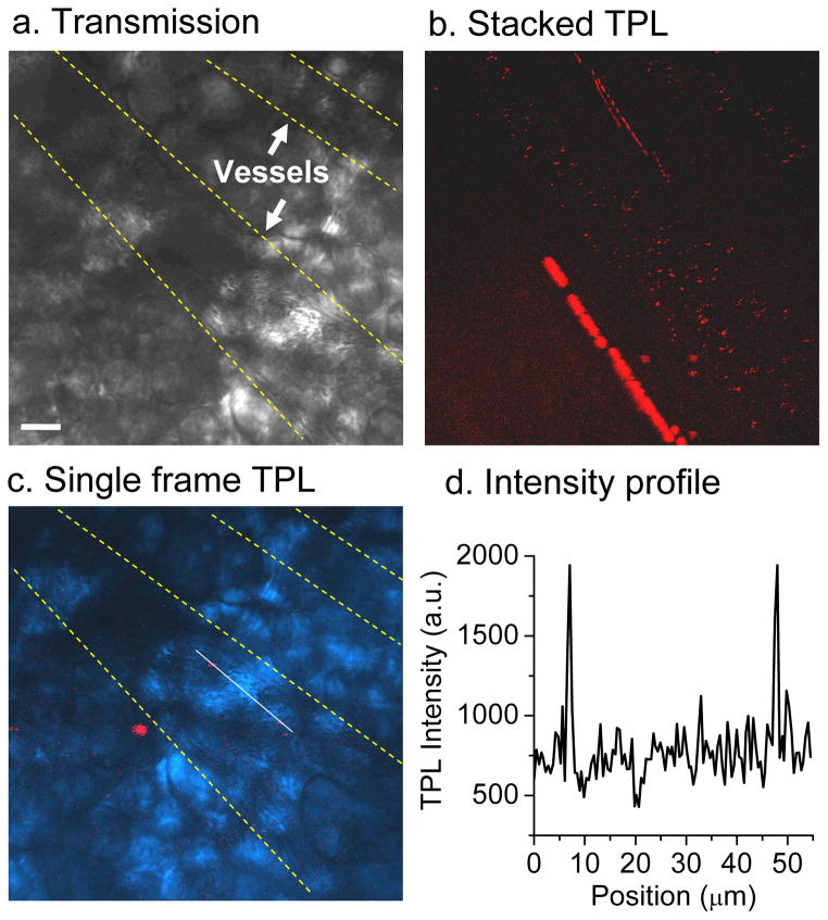
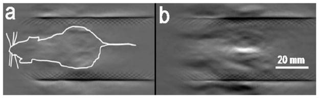
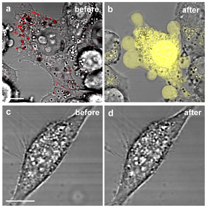
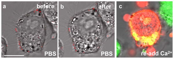
Gold nanorods (NRs) have plasmon-resonant absorption and scattering in the near-infrared (NIR) region, making them attractive probes for in vitro and in vivo imaging. In the cellular environment, NRs can provide scattering contrast for darkfield microscopy, or emit a strong two-photon luminescence due to plasmon-enhanced two-photon absorption. NRs have also been employed in biomedical imaging modalities such as optical coherence tomography or photoacoustic tomography. Careful control over surface chemistry enhances the capacity of NRs as biological imaging agents by enabling cell-specific targeting, and by increasing their dispersion stability and circulation lifetimes. NRs can also efficiently convert optical energy into heat, and inflict localized damage to tumor cells. Laser-induced heating of NRs can disrupt cell membrane integrity and homeostasis, resulting in Ca(2+) influx and the depolymerization of the intracellular actin network. The combination of plasmon-resonant optical properties, intense local photothermal effects and robust surface chemistry render gold NRs as promising theragnostic agents.


Figures

Figure 1
Figure 1. Photophysical processes in gold NRs
NIR irradiation induces the excitation of a LPR mode, resulting mostly in absorption but also some resonant light scattering. An electronic transition from the d- band to sp- band occurs with two-photon absorption, generating an electron-hole pair; recombination of separated charges results in TPL emission. Heat is also generated as a consequence of electron-phonon collisions.
Figure 2
Figure 2. Gold NRs with tunable optical absorptions at visible and NIR wavelengths
(a) Optical absorption spectra of gold NRs with different aspect ratios. (b) Color wheel, with reference to λLR and λTR for gold NRs labeled ae.
Figure 3
Figure 3. Surface functionalization of gold NRs
(a) Electrostatic adsorption onto polyelectrolyte-coated gold NRs. (b) Conjugation of amine-terminated biotin by carbodiimide coupling (102). (c) Cu-catalyzed “click” addition of alkyne-terminated molecules onto azide-labeled surface (103). (d) Chemisorption of thiolated bioconjugates (48, 53, 59). (e) Chemisorption of amine-terminated bioconjugates by in situ dithiocarbamate (DTC) formation (33, 34, 45, 57).
Figure 4
Figure 4. Characterization of TPL from gold NRs by far-field microscopy
(a) A typical TPL spectrum from a single NR excited by fs-pulsed laser centered at its LPR peak at 786 nm. The laser power was 1.5 mW and acquisition time was 1 s. The spectrum was cut off at 650 nm by an IR blocker. (b) Quadratic dependence of TPL emission intensity on excitation power. The data were obtained by increasing the excitation pulse energy from 1 to 2 pJ, then decreasing the power accordingly. Signal intensities were integrated over an area of 250 × 250 μm (18). (c) TPL excitation spectrum (circles with error bars, right y-axis) compared with NR extinction. The peak at 820 nm corresponds to the longitudinal plasmon mode. The same power (0.17 mW) was used for all excitation wavelengths (18). (d) TPL spectra of three NR solutions with resonance peaks at 660 nm (black line), 786 nm (red line), and 804 nm (green line), irradiated by the same excitation laser (785 nm, 3 mW; acquisition time 60 s). TPL spectra were cut off at 650 nm by an IR blocker.
Figure 5
Figure 5. Polarization-dependent TPL intensities of gold NRs
(18). (a,b) Pseudocolor images with horizontal (θ=0°) and vertical (θ=90°) excitation polarizations, respectively. Single NRs could be identified by their similar range of intensities (e.g. spots 1, 2); brighter spots were considered to be clusters of NRs (e.g. spot 3). (b) Polarization dependence of the TPL intensity (solid dots) for a single NR (spot 2). The excitation polarization was rotated clockwise from −90° to +90° in 10° increments. The TPL signal fits a cos4 function, offset by 6.8° (red curve). (d) The TPL emission (solid dots) from the same NR (spot 2) measured by rotating a polarizer before the detector. The reflected excitation beam was linearly polarized at the detector site (solid curve), but the TPL emission was essentially depolarized.
Figure 6
Figure 6. In vitro cellular imaging with F-NRs (34)
(a) folate-oligoethyleneglycol ligands, conjugated onto NR surface by in situ dithiocarbamate formation. (b) F-NRs bound to the KB cell surface after incubation for 6 hours. (c) Almost no F-NRs were observed to be associated with NIH/3T3 cells, which do not overexpress the high-affinity folate receptor. (d) F-NRs were internalized into KB cells and delivered to the perinuclear area after incubation for 17 hours. (e) TPL intensity profile across green line in (d). Bar= 10 μm.
Figure 7
Figure 7. In vivo TPL imaging of NRs in blood vessels
(18). (a) Transmission image with two blood vessels indicated. (b) TPL image of CTAB-NRs (red dots) flowing through blood vessels. The image was compiled by stacking 300 frames collected continuously at a rate of 1.12 s per frame. (c) Overlay of transmission image (blue) and a single-frame TPL image. Two NRs (red) are superimposed by a linescan. (d) TPL intensity profile from the linescan in (c). Bar = 20 μm.
Figure 8
Figure 8. NRs as contrast agents for photoacoustic tomography
(88). (a) PAT image of a nude mouse (white outline) prior to injection of NRs; (b) PAT contrast after NR injection. Reprinted with permission from the American Chemical Society.
Figure 9
Figure 9. Photothermolysis mediated by F-NRs
(34). (a,b) KB cells with membrane-bound F-NRs (red) exposed to fs-pulsed NIR laser irradiation (0.75 mW, 81.4 s) experienced membrane damage and blebbing. The loss of membrane integrity was indicated by ethidium bromide (EB) nuclear staining (yellow). (c,d) NIH-3T3 cells were unresponsive to F-NRs and did not suffer photoinduced damage at the same condition. Bar= 10 μm.
Figure 10
Figure 10. Ca2+-dependent membrane blebbing during nanorod-mediated photothermolysis (34)
(a–b) No blebbing was observed for F-NRs labeled KB cells in Ca2+-free PBS after fs-pulsed laser irradiation at 3 mW for 61.5 s. (c) Blebs were immediately produced upon addition of 0.9 mM Ca2+. Incubation with 2.5 μM EB (red) and 2 μM Oregon Green for 20 min indicated a compromise in membrane integrity and an elevation in intracellular Ca2+. For all experiments, cells were incubated with F-NRs for 6 h, and washed 5 times in Ca2+-free PBS. Amounts of dyes and reagents were described as final concentrations in the cell culture medium. Bar = 10 μm.

References

    1. Murphy CJ, Sau TK, Gole AM, Orendorff CJ, Gao J, Gou L, Hunyadi SE, Li T. Anisotropic metal nanoparticles: synthesis, assembly, and optical applications. J Phys Chem B. 2005;109:13857–13870. - PubMed
    1. Pérez-Juste J, Pastoriza-Santos I, Liz-Marzán LM, Mulvaney P. Gold nanorods: synthesis, characterization and applications. Coord Chem Rev. 2005;249:1870–1901.
    1. Hirsch LR, Gobin AM, Lowery AR, Tam F, Drezek RA, Halas NJ, West JL. Metal nanoshells. Annals Biomed Eng. 2006;34:15–22. - PubMed
    1. Huang X, Jain PK, El-Sayed IH, El-Sayed MA. Gold nanoparticles: interesting optical properties and recent applications in cancer diagnostics and therapy. Nanomed. 2007;2:681–693. - PubMed
    1. Skrabalak SE, Chen J, Sun Y, Lu X, Au L, Cobley CM, Xia Y. Gold nanocages: synthesis, properties, and applications. Acc Chem Res. 2008 in press. - PMC - PubMed

Publication types

LinkOut - more resources