. 2009 Apr;86(2):87-94.
doi: 10.1016/j.yexmp.2008.12.005. Epub 2009 Jan 7.

Response gene to complement 32 is required for C5b-9 induced cell cycle activation in endothelial cells

Affiliations

Response gene to complement 32 is required for C5b-9 induced cell cycle activation in endothelial cells

Matthew Fosbrink et al. Exp Mol Pathol. 2009 Apr.

Abstract

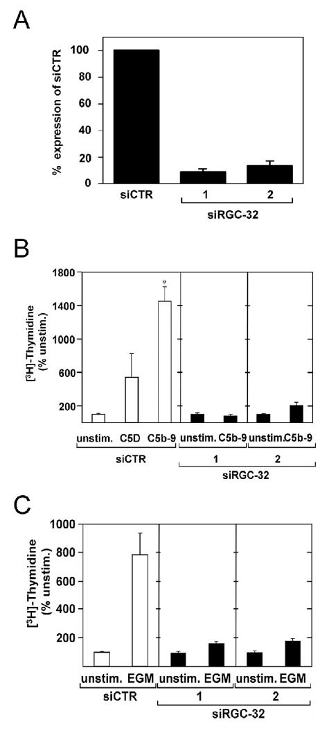
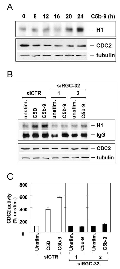
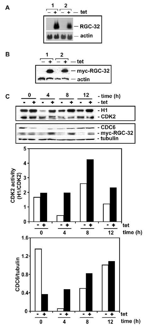
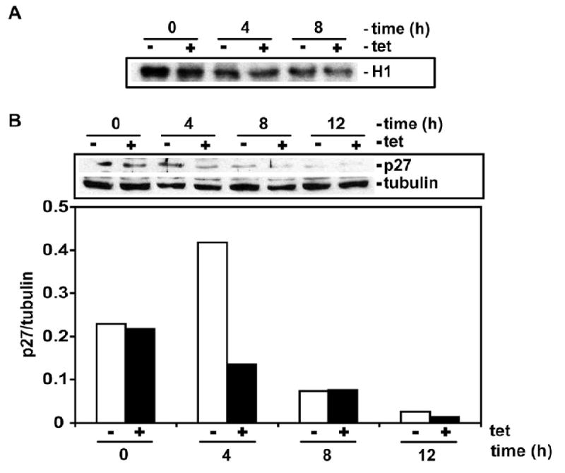
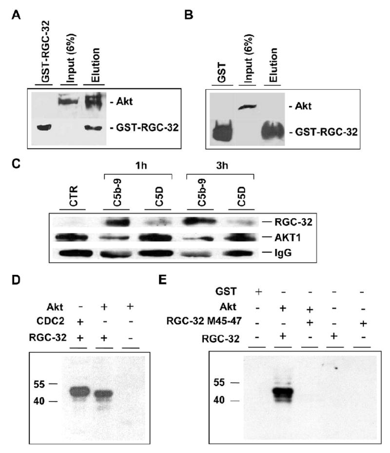
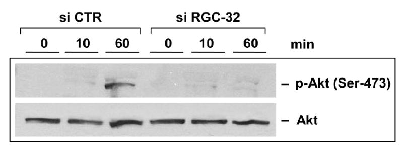
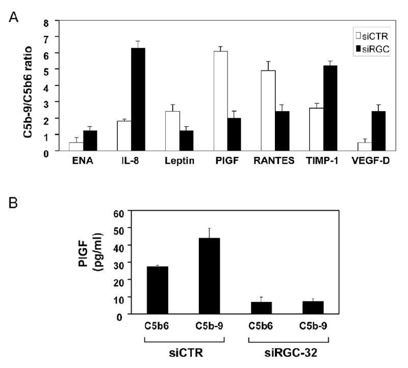
Proliferation of vascular endothelial cells (EC) and smooth muscle cells (SMC) is a critical event in angiogenesis and atherosclerosis. We previously showed that the C5b-9 assembly during complement activation induces cell cycle in human aortic EC (AEC) and SMC. C5b-9 can induce the expression of Response Gene to Complement (RGC)-32 and over expression of this gene leads to cell cycle activation. Therefore, the present study was carried out to test the requirement of endogenous RGC-32 for the cell cycle activation induced by C5b-9 by knocking-down its expression using siRNA. We identified two RGC-32 siRNAs that can markedly reduce the expression of RGC-32 mRNA in AEC. RGC-32 silencing in these cells abolished DNA synthesis induced by C5b-9 and serum growth factors, indicating the requirement of RGC-32 activity for S-phase entry. RGC-32 siRNA knockdown also significantly reduced the C5b-9 induced CDC2 activation and Akt phosphorylation. CDC2 does not play a role in G1/S transition in HeLa cells stably overexpressing RGC-32. RGC-32 was found to physically associate with Akt and was phosphorylated by Akt in vitro. Mutation of RGC-32 protein at Ser 45 and Ser 47 prevented Akt mediated phosphorylation. In addition, RGC-32 was found to regulate the release of growth factors from AEC. All these data together suggest that cell cycle induction by C5b-9 in AEC is RGC-32 dependent and this is in part through regulation of Akt and growth factor release.


Figures

Figure 1
Figure 1. RGC-32 is required for C5b-9 and growth factors induced cell cycle
A. AEC were transfected with siRGC-32-1, siRGC-32–2, or control siRNA (siCTR) at a final concentration of 25 nM using Mirus SiQuest transfection reagent. After 48 h, total RNA was extracted, and analyzed for RGC-32 and Actin mRNA expression by real time - PCR. Both siRGC-32 significantly reduced RGC-32 mRNA expression. B. AEC were stimulated with serum C5b-9 (C5b-9) or C5D serum for 24 h in the presence of 1 μCi [3H]-thymidine. The level of [3H]-thymidine incorporation was used to assess cell cycle activation. C5b-9 significantly increased [3H]-thymidine incorporation compared to C5D (*p<0.001). Both siRGC-32-1 and −2 abolished thymidine incorporation induced by C5b-9. C. AEC were transfected with siRGC-32-1, siRGC-32-2, or control siCTR for 48 h. Cells were starved in serum- and growth factor-free medium for 18 h to synchronize in G0/G1-phase of the cell cycle. Cells were then stimulated with endothelial growth media (EGM) for 24 h, followed by [3H]-thymidine incorporation. Both RGC-32 siRNAs abolished EGM-induced [3H]-thymidine incorporation. Results are expressed as % of unstimulated cell level (Unstim).
Figure 2
Figure 2. RGC-32 is required for the C5b-9 induced CDC2 activation
A. AEC were cultured in serum- and growth factor-free medium for 18 h, then exposed to serum C5b-9 for the times indicated. CDC2 was then immunoprecipitated and the activity was assayed using histone H1 as a substrate in the presence of γ32P-ATP (upper panel – H1) followed by SDS-PAGE and transfer to nitrocellulose membrane. Autoradiography revealed phosphorylated histone H1 bands, representing CDC2 activity. The CDC2 and tubulin protein levels were determined by western blot using anti-CDC2 IgG (lower panel). C5b-9 increased CDC2 activity in a time-dependent manner without any significant changes in the CDC2 and tubulin protein level. B. siRGC-32-1, siRGC-32-2, or control siCTR were complexed with Mirus siQuest transfection reagent, and added to EC, at a final concentration of 25 nM. After 48 h, cells were starved in serum- and growth-factor-free medium for 18 h and then stimulated with serum C5b-9 (C5b-9) or C5D for 24 h. Cells were lysed and CDC2 activity was assayed (upper panel – H1). The IgG levels were used as loading control. The same blot was probed with anti-CDC2 IgG and tubulin by western blotting. C. The CDC2 activity was quantitated by densitometric scanning of phosphorylated histone H1 bands and by normalizing the values to IgG. CDC2 was expressed as % of unstimulated cell activity (n=3). Significantly higher levels of CDC2 activity were induced by C5b-9, when compared to the level of unstimulated- or C5D-stimulated cells. The CDC2 activity induced by C5b-9 was abolished in cells transfected with both RGC-32 siRNAs.
Figure 3
Figure 3. Effect of RGC-32 overexpression on CDK2 and CDC6 activity
A, B. HeLa cell line stably overexpressing RGC-32. Two clones were examined for tetracycline induction of RGC-32 expression by northern blotting (A) and western blotting (B). For western blotting we used an antibody that recognizes the Myc-tag that is fused to the C-terminal of RGC-32. RGC-32 expression is induced by exposure to tetracycline (2 μg/ml). C. RGC-32 accelerates and enhances CDK2 and CDC6 activity. HeLa cells overexpressing RGC-32 were arrested in M-phase by nocodazole block and released into growth media in the presence (+) or absence (-) of 2 μg/ml of tetracycline (tet). At the times indicated, cells were collected and lysed. 0 h post-M represents cells that were released from nocodazole blocked then immediately lysed. For CDK2 kinase assay (upper panel) immunoprecipitated CDK2 was incubated with [γ-32P] ATP and histone H1. Phosphorylated histone H1 (upper panel - H1) was detected by autoradiography. The same blot was used to detect CDK2 by western blotting (upper panel – CDK2). Whole lysates were used to detect CDC6, myc-RGC-32 and tubulin expression. To quantitate CDK2 activity and CDC6 densitometric scanning was performed.
Figure 4
Figure 4. Effect of RGC-32 overexpression on CDC2 activity and p27 expression
HeLa RGC-32 overexpressing were arrested in M-phase by nocodazole block and released into growth media in the presence (+) or absence (-) of 2 μg/ml of tetracycline (tet). At the times indicated, cells were collected and lysed. 0 h post-M represents cells that were released from nocodazole blocked then immediately lysed. For CDC2 kinase assay (upper panel) immunoprecipitated CDC2 was incubated with [γ-32P] ATP and histone H1. Phosphorylated histone H1 (upper panel - H1) was detected by autoradiography. Whole lysates was used to detect p27 and tubulin expression.
Figure 5
Figure 5. RGC-32 interacts and regulates Akt
A, B. Formation of RGC-32-Akt complexes. GST-RGC-32 fusion protein (A) or GST (negative control) (B) interaction with Akt in AEC lysates was analyzed using the Profound pull-down GST protein-protein interaction kit (Pierce). The proteins binding to GST-RGC-32 or GST in the presence of glutathione-Sepharose beads were eluted and analyzed by Western blotting using anti-Akt antibodies. A. Akt was eluted together with GST-RGC-32 (A; lane Elution). Akt input is also shown (A; Input 6% lane). B. Akt was not recovered with GST alone (B; lane GST). Akt input is shown (B; Input 6% lane). C. Coimmunoprecipitation of RGC-32 with Akt in vivo. AEC treated with C5b-9 or C5b6 (C5D) for 1 and 3 h were lysed and immunoprecipitated with anti-Akt IgG in the presence of protein A/G-agarose. Protein eluted from the beads separated in 10% SDS-PAGE and then immunoblotted with anti-RGC-32 and anti-Akt. IgG was used for normalization. CTR., unstimulated cells. D, E. Phosphorylation of RGC-32 by Akt in vitro. Phosphorylation of RGC-32 by Akt was examined by in vitro kinase assay. Recombinant RGC-32-GST (1 μM) or GST (1 μM) were incubated with Akt (1μM) in the presence of 1 μCi of [γ-32P] ATP/sample as described in Material and Methods section. The phosphorylation of GST-RGC-32 was assessed by SDS-PAGE and then autoradiography. GST-RGC-32 (revealed as a 40 kD band) but not GST was phosphorylated by Akt (D). Phosphorylation of GST- RGC-32 by CDC2/cyclin B1 was used as a positive control (Badea et al., 2002). Mutation of Serine 45 and Seine 47 in RGC-32 molecule resulted in the abolition of GST-RGC-32 phosphorylation by Akt (E).
Figure 6
Figure 6. RGC-32 is required for C5b-9-induced Akt phosphorylation
AEC were transfected with RGC-32 siRNA or control CTR siRNA, starved in serum- and growth factor-free media for 18 h, then stimulated with serum C5b-9, as described earlier, for 10 and 60 min. Cells were lysed and Akt phosphorylation was assessed by western blot, using anti-Akt IgG specific for Akt phosphorylated at Ser 473. RGC-32 knockdown results in inhibition of Akt phosphorylation by C5b-9.
Figure 7
Figure 7. RGC-32 is required for the release of angiogenic factors by C5b-9 and mediation of PIGF release
A. AEC were transfected with siRGC-32-1, or control siCTR, then starved in serum- and growth factor-free media for 18 h. Cells were then stimulated with C5b6 or C5b-9 assembled from purified components for 1 h. The conditioned media were subjected to an angiogenic factor antibody protein array. The array was quantitated by densitometric scanning and C5b-9/C5b6 ratios were calculated. Only those factors exhibiting a 1.5-fold difference in C5b-9/C5b6 ratio are shown. B. AEC transfected with siRGC-32 or control siCTR and cultured in serum and growth factor free media, as described, were stimulated with C5b6 or C5b-9 assembled from purified components for 1 h. The conditioned media were tested for the presence of PlGF by ELISA. PlGF release was inhibited in cells transfected with siRGC-32.

Similar articles

Cited by

References

    1. Badea T, Niculescu F, Soane L, Fosbrink M, Sorana H, Rus V, Shin ML, Rus H. RGC-32 increases p34CDC2 kinase activity and entry of aortic smooth muscle cells into S-phase. J Biol Chem. 2002;277:502–8. - PubMed
    1. Badea TC, Niculescu FI, Soane L, Shin ML, Rus H. Molecular cloning and characterization of RGC-32, a novel gene induced by complement activation in oligodendrocytes. J Biol Chem. 1998;273:26977–81. - PubMed
    1. Bazzoni G, Dejana E. Endothelial cell-to-cell junctions: Molecular organization and role in vascular homeostasis. Physiol Rev. 2004;84:869–901. - PubMed
    1. Benzaquen LR, Nicholson-Weller A, Halperin JA. Terminal complement proteins C5b-9 release basic fibroblast growth factor and platelet-derived growth factor from endothelial cells. J Exp Med. 1994;179:985–92. - PMC - PubMed
    1. Carney DF, Koski CL, Shin ML. Elimination of terminal complement intermediates from the plasma membrane of nucleated cells: the rate of disappearance differs for cells carrying C5b-7 or C5b-8 or a mixture of C5b-8 with a limited number of C5b-9. J Immunol. 1985;134:1804–9. - PubMed

Publication types

MeSH terms