. 2009 Mar 6;386(4):938-50.
doi: 10.1016/j.jmb.2008.12.080. Epub 2009 Jan 8.

Hepatoma-derived growth factor represses SET and MYND domain containing 1 gene expression through interaction with C-terminal binding protein

Affiliations

Hepatoma-derived growth factor represses SET and MYND domain containing 1 gene expression through interaction with C-terminal binding protein

Jun Yang et al. J Mol Biol. .

Abstract

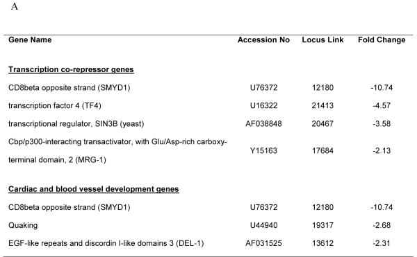
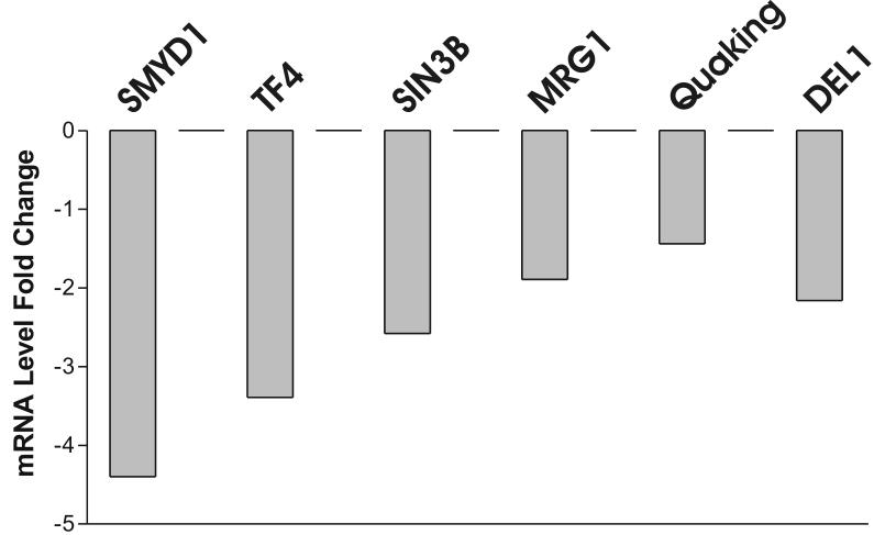
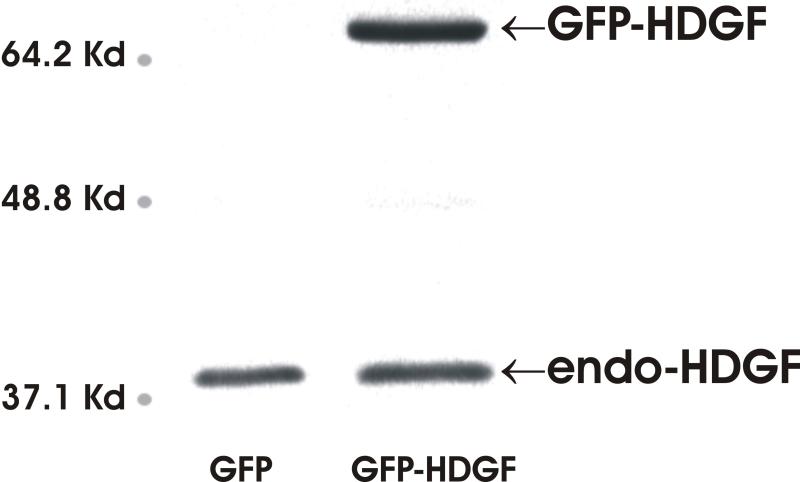
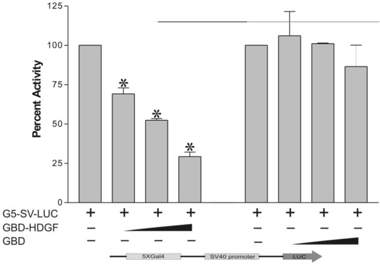
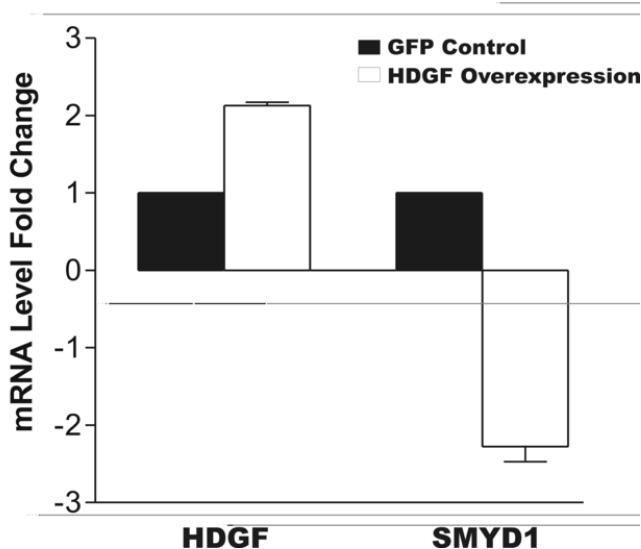
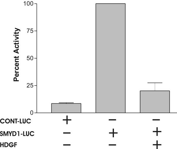
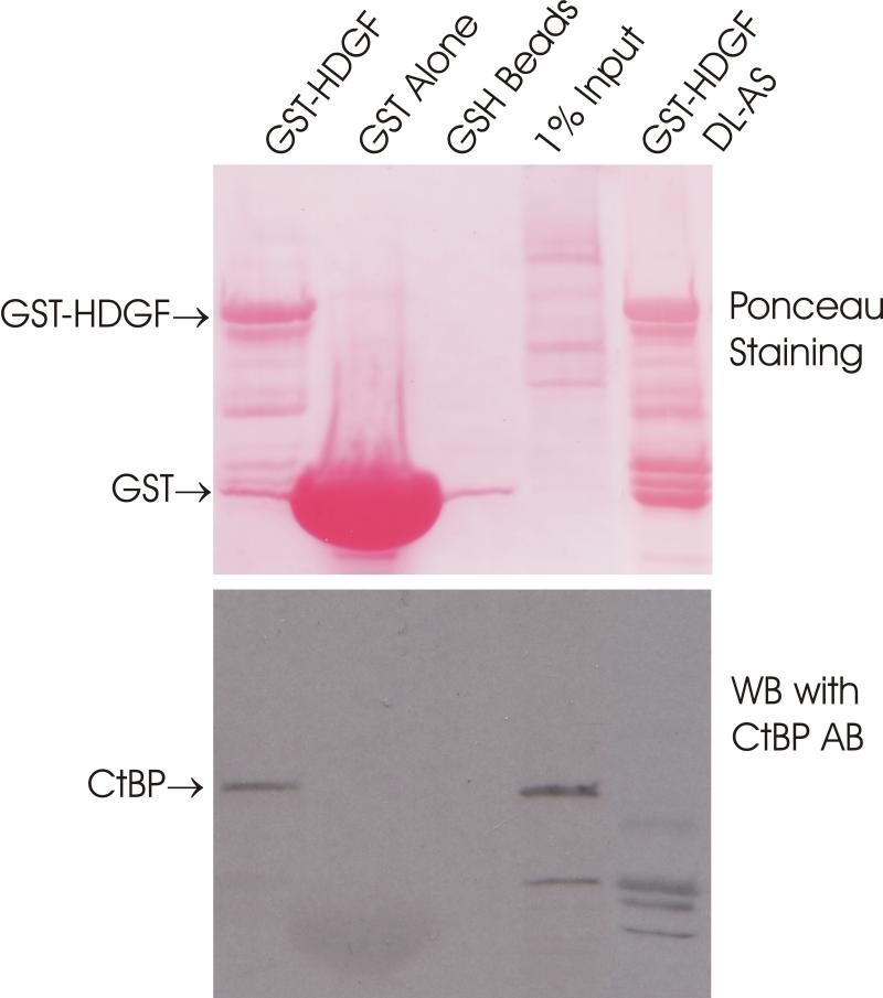
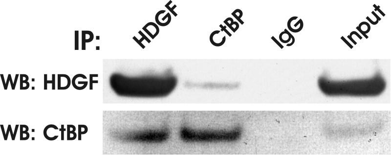
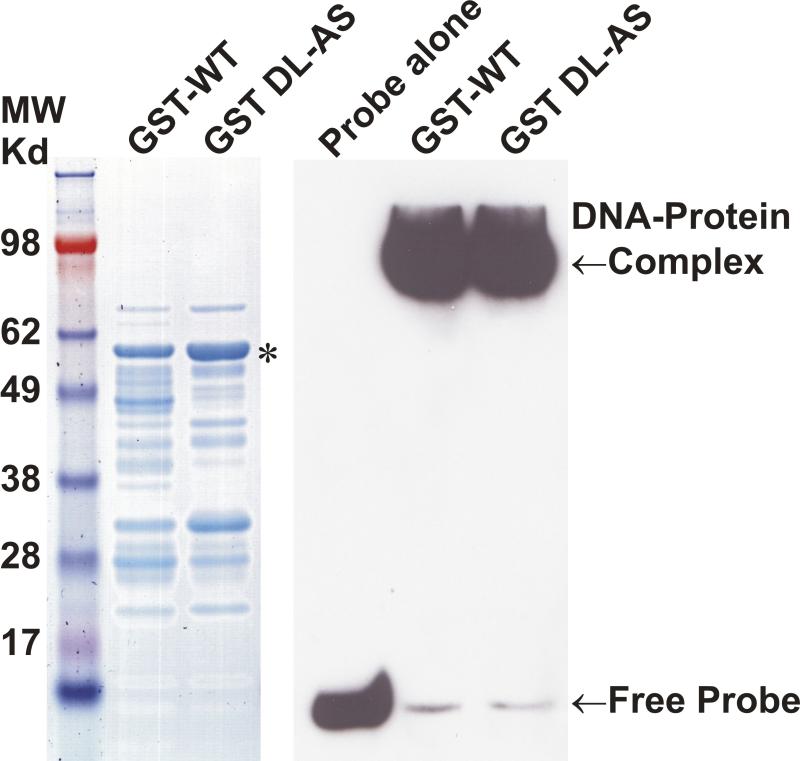
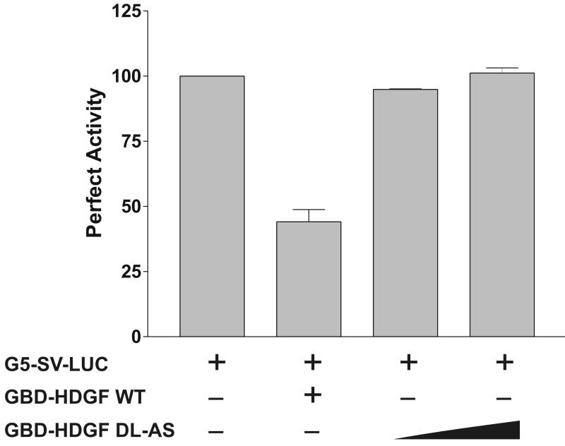
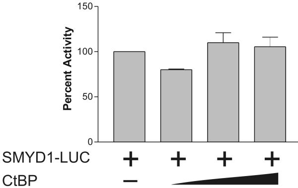
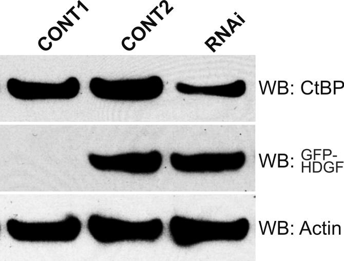
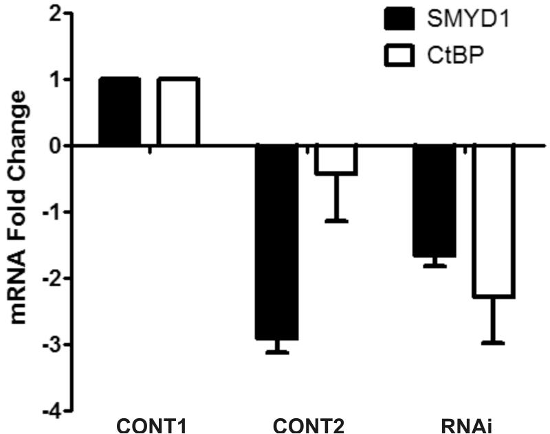
Hepatoma-derived growth factor (HDGF) is a nuclear protein with both mitogenic and angiogenic activity that is highly expressed in the developing heart and vasculature. To date, the mechanism underlying the function of HDGF is unknown. Oligonucleotide microarray analysis was used to gain insights into HDGF function. Adenoviral expression of HDGF significantly (> or =2-fold) downregulated a large group (66) of genes, and increased expression of a relatively small number of genes (9). Two groups of target genes that are involved in cardiovascular development and transcriptional regulation, including the skeletal/cardiac muscle specific SET and MYND domain containing 1 (SMYD1) gene, were validated by real time PCR. This suggested that HDGF could function as a transcriptional repressor. In a one-hybrid system, GBD-HDGF significantly repressed reporter gene activity in a dose-dependent manner. This demonstrated that HDGF has transcriptional repressive activity. Moreover, in G-7 myoblast cells, over-expression of a GFP-HDGF fusion specifically downregulated SMYD1 mRNA expression and the activity of the human SMYD1 promoter. HDGF repressed SMYD1 gene transcription through interaction with a transcriptional corepressor C-terminal binding protein (CtBP). Over-expression of CtBP potentiated the trans-repressive activity of HDGF; on the other hand, knocking down CtBP attenuated the trans-repressive effect of HDGF. HDGF binds CtBP through a non-canonical binding motif (PKDLF) within the PWWP domain, as mutation of DL to AS abolished HDGF and CtBP interaction, and diminished the trans-repressive effect of HDGF without affecting DNA binding. Finally, fluorescent microscopy showed that HDGF induced the nuclear accumulation of CtBP, suggesting that HDGF forms a transcriptional complex with CtBP. Taken together, our data demonstrate that HDGF functions as a transcriptional repressor of the SMYD1 gene through interaction with the transcriptional corepressor CtBP. Because of moderate conservation of the CtBP binding motif in HDGF family members, trans-repressive activity mediated by CtBP may be a common function among HDGF proteins.


Figures

Figure 1
Figure 1
Representative genes downregulated by HDGF. (A) Classified microarray results shown that a group of 4 genes serve as transcription co-repressor and a group of 3 involved in cardiac and blood vessel development. (B) Real Time PCR confirmed the microarray result. Mouse smooth muscle cells were infected with GFP-HDGF adenovirus and GFP virus as control for 18 hours, total RNA was extracted and reverse transcripted. Real Time PCR analysis was performed using gene specific primers for 7 genes listed in Table 1. Bar graph shows mRNA levels change. (C) Western blot using HDGF polyclonal antibody shows the expression of endogenous HDGF and GFP-HDGF fusion.
Figure 1
Figure 1
Representative genes downregulated by HDGF. (A) Classified microarray results shown that a group of 4 genes serve as transcription co-repressor and a group of 3 involved in cardiac and blood vessel development. (B) Real Time PCR confirmed the microarray result. Mouse smooth muscle cells were infected with GFP-HDGF adenovirus and GFP virus as control for 18 hours, total RNA was extracted and reverse transcripted. Real Time PCR analysis was performed using gene specific primers for 7 genes listed in Table 1. Bar graph shows mRNA levels change. (C) Western blot using HDGF polyclonal antibody shows the expression of endogenous HDGF and GFP-HDGF fusion.
Figure 2
Figure 2
HDGF represses the expression of a one-hybrid reporter gene. The reporter gene G5-SV-LUC (Firefly luciferase 0.5 ug), pRL-CMV (Renilla luciferase 50 ng) and GBD-HDGF (0.5, 1, and 2 ug in left panel), or GBD (0.5, 1 and 2 ug in right panel) alone were cotransfected into G-7 cells. The dual-luciferase reporter assay system was used to measure the luciferase activities 24 hours after transfection. Percentage of firefly luciferase activity was calculated when luciferase activity with reporter gene alone was set at 100%. Firefly luciferase activity was normalized with constitutive renilla luciferase activity to correct transfection efficiency. Bars indicate mean ± SEM from 3 separate transfection assays with duplicate plates, *P < 0.05 by one-way ANOVA control versus GBD-HDGF.
Figure 3
Figure 3
HDGF represses the expression of SMYD1 gene. A, G-7 cells were infected with 100 MOI adenovirus expressing GFP-HDGF fusion or GFP for 18 hours, total RNA were extracted and reverse transcribed for real-time PCR analysis. Gene specific primers of HDGF and SMYD1 were used to detect their mRNA levels. Specific primers amplifying the mouse TATA Box Binding Protein (TBP) were used to normalize gene expression levels. Bars indicate mean ± SEM from 2 separate experiments with triplicate assays. B, 1 ug pGL3-enhancer vector as CONT-LUC or SMYD1 promoter reporter SMYD1-LUC were cotransfected with 50 ng pRL-CMV with or without 3ug HDGF expression construct into G-7 cells. The dual-luciferase reporter assay system was used to measure the luciferase activities 24 hours after transfection. Percentage of firefly luciferase activity was calculated when SMYD1-LUC luciferase activity without HDGF was set at 100%. Firefly luciferase activity was normalized with constitutive renilla luciferase activity to correct transfection efficiency. Bars indicate mean ± SEM from 3 separate transfection assays with duplicate plates.
Figure 3
Figure 3
HDGF represses the expression of SMYD1 gene. A, G-7 cells were infected with 100 MOI adenovirus expressing GFP-HDGF fusion or GFP for 18 hours, total RNA were extracted and reverse transcribed for real-time PCR analysis. Gene specific primers of HDGF and SMYD1 were used to detect their mRNA levels. Specific primers amplifying the mouse TATA Box Binding Protein (TBP) were used to normalize gene expression levels. Bars indicate mean ± SEM from 2 separate experiments with triplicate assays. B, 1 ug pGL3-enhancer vector as CONT-LUC or SMYD1 promoter reporter SMYD1-LUC were cotransfected with 50 ng pRL-CMV with or without 3ug HDGF expression construct into G-7 cells. The dual-luciferase reporter assay system was used to measure the luciferase activities 24 hours after transfection. Percentage of firefly luciferase activity was calculated when SMYD1-LUC luciferase activity without HDGF was set at 100%. Firefly luciferase activity was normalized with constitutive renilla luciferase activity to correct transfection efficiency. Bars indicate mean ± SEM from 3 separate transfection assays with duplicate plates.
Figure 4
Figure 4
HDGF interacted with CtBP. A, GST-HDGF fusion proteins (lane 1, wild type, lane 5, DL-AS mutant) were conjugated to glutathione-agarose beads and incubated with whole cell lysate of MCF-7 cells, as described under “Experimental Procedures.” After washing and recovery of the beads, associated proteins were resolved by SDS-PAGE and analyzed by western blot with anti-CtBP antibody. Top panel shows Ponceau staining of the nitrocellulose membrane. B, Endogenous HDGF or CtBP were immunoprecipitated from MCF-7 cell lysates with either monoclonal anti-HDGF antibody, or monoclonal anti-CtBP antibody, the immunoprecipitated protein complexes were detected by immunoblotting with Rabbit polyclonal anti-HDGF or anti-CtBP antibodies. C, Flag-CtBP was co-expressed with either HA-HDGF wild type or DL-AS mutant form in MCF-7 cells, co-immunoprecipitation were done by either IP with HA antibody, western blotting with Flag antibody, or IP with Flag antibody, western blotting with HA antibody. Western blotting with HA and Flag antibodies for whole cell lysate were also shown. D, 62DL-AS mutant HDGF binds to DNA. 0.25 pmol 5′ end biotin labeled probes for SMYD1 promoter (nucleotides -688 to -609) were incubated with 1 ug GST-HDGF fusion protein (wild type or 62DL-AS mutant) in the reaction buffer on ice for 30 min. The complex were resolved in 5% PAGE and transferred onto a nylon membrane. The biotin end-labeled DNA probe is detected using a streptavidin-horseradish peroxidase conjugate and chemiluminescent substrate. The left panel shows the coomassie staining of the fusion proteins, star indicates the full length of HDGF fusion proteins. E, 62DL-AS mutant does not have the trans-repressive activity. Wild type (2ug) or 62DL-AS mutant forms of GBD-HDGF expression constructs (1 and 2 ug) were cotransfected with G5-SV-LUC (0.5 ug) and pRL-CMV (50 ng), dual-luciferase assay was performed as described in Figure 2.
Figure 4
Figure 4
HDGF interacted with CtBP. A, GST-HDGF fusion proteins (lane 1, wild type, lane 5, DL-AS mutant) were conjugated to glutathione-agarose beads and incubated with whole cell lysate of MCF-7 cells, as described under “Experimental Procedures.” After washing and recovery of the beads, associated proteins were resolved by SDS-PAGE and analyzed by western blot with anti-CtBP antibody. Top panel shows Ponceau staining of the nitrocellulose membrane. B, Endogenous HDGF or CtBP were immunoprecipitated from MCF-7 cell lysates with either monoclonal anti-HDGF antibody, or monoclonal anti-CtBP antibody, the immunoprecipitated protein complexes were detected by immunoblotting with Rabbit polyclonal anti-HDGF or anti-CtBP antibodies. C, Flag-CtBP was co-expressed with either HA-HDGF wild type or DL-AS mutant form in MCF-7 cells, co-immunoprecipitation were done by either IP with HA antibody, western blotting with Flag antibody, or IP with Flag antibody, western blotting with HA antibody. Western blotting with HA and Flag antibodies for whole cell lysate were also shown. D, 62DL-AS mutant HDGF binds to DNA. 0.25 pmol 5′ end biotin labeled probes for SMYD1 promoter (nucleotides -688 to -609) were incubated with 1 ug GST-HDGF fusion protein (wild type or 62DL-AS mutant) in the reaction buffer on ice for 30 min. The complex were resolved in 5% PAGE and transferred onto a nylon membrane. The biotin end-labeled DNA probe is detected using a streptavidin-horseradish peroxidase conjugate and chemiluminescent substrate. The left panel shows the coomassie staining of the fusion proteins, star indicates the full length of HDGF fusion proteins. E, 62DL-AS mutant does not have the trans-repressive activity. Wild type (2ug) or 62DL-AS mutant forms of GBD-HDGF expression constructs (1 and 2 ug) were cotransfected with G5-SV-LUC (0.5 ug) and pRL-CMV (50 ng), dual-luciferase assay was performed as described in Figure 2.
Figure 5
Figure 5
CtBP mediated the trans-repressive activity of HDGF. A, Reporter gene construct G5-SV-LUC (0.5 ug) and pRL-CMV (50 ng) were cotransfected with (left panel) or without GBD-HDGF (right panel) and increasing amounts of CtBP expression construct (0.5, 1, 2 ug) as indicated in the figure in G7 cells, the dual-luciferase assay was performed as described in Fig 2. *P < 0.05 by one-way ANOVA control versus CtBP (Left panel). B, CtBP alone does not repress SMYD1 gene promoter activity. SMYD1 promoter reporter SMYD1-LUC (1ug) was cotransfected with 50 ng pRL-CMV and increasing amount of CtBP expression construct (1, 2, 3 ug) into G-7 cells. The dual-luciferase reporter assay was performed as described in Fig 3. C, HDGF expression constructs (2ug) were cotransfected with 0, 3 or 4 ug Flag-CtBP in G7 cells for 48 hours, real-time PCR were performed as described in Fig 3 to measure the mRNA levels change of SMYD1 gene. D, 1 ug GFP-HDGF was cotransfected with 1 ug of either conrol or specific miR targeting CtBP expression vectors into MCF-7 cells which cultured in 6 wells plate using lipofectamine 2000 reagent. 72 hours after transfection, the protein lysate were subjected to SDS-PAGE and further western blotting using specific antibodies against CtBP, GFP or Actin as control. Control 1 used empty GFP vector instead of GFP-HDGF. E, Total RNA extracted from MCF-7 cells described in 4D was subjected to real-time PCR. The mRNA levels of both SMYD1 and CtBP in control 1 (GFP vector plus control miR vector) were set to 1.
Figure 5
Figure 5
CtBP mediated the trans-repressive activity of HDGF. A, Reporter gene construct G5-SV-LUC (0.5 ug) and pRL-CMV (50 ng) were cotransfected with (left panel) or without GBD-HDGF (right panel) and increasing amounts of CtBP expression construct (0.5, 1, 2 ug) as indicated in the figure in G7 cells, the dual-luciferase assay was performed as described in Fig 2. *P < 0.05 by one-way ANOVA control versus CtBP (Left panel). B, CtBP alone does not repress SMYD1 gene promoter activity. SMYD1 promoter reporter SMYD1-LUC (1ug) was cotransfected with 50 ng pRL-CMV and increasing amount of CtBP expression construct (1, 2, 3 ug) into G-7 cells. The dual-luciferase reporter assay was performed as described in Fig 3. C, HDGF expression constructs (2ug) were cotransfected with 0, 3 or 4 ug Flag-CtBP in G7 cells for 48 hours, real-time PCR were performed as described in Fig 3 to measure the mRNA levels change of SMYD1 gene. D, 1 ug GFP-HDGF was cotransfected with 1 ug of either conrol or specific miR targeting CtBP expression vectors into MCF-7 cells which cultured in 6 wells plate using lipofectamine 2000 reagent. 72 hours after transfection, the protein lysate were subjected to SDS-PAGE and further western blotting using specific antibodies against CtBP, GFP or Actin as control. Control 1 used empty GFP vector instead of GFP-HDGF. E, Total RNA extracted from MCF-7 cells described in 4D was subjected to real-time PCR. The mRNA levels of both SMYD1 and CtBP in control 1 (GFP vector plus control miR vector) were set to 1.
Figure 6
Figure 6
HDGF induced the nuclear accumulation of CtBP. MCF-7 cells grown on cover slips were transfected with 0.5 ug of CFP-CtBP and GFP-HDGF (top panel) or GFP (2nd panel) for 48 hours, cells were fixed in 1% paraformaldehyde and the cover slips were mounted in DAPI containing medium, DAPI, CFP and GFP images are shown, with the merged images of CFP and GFP fluorescence (overlay). MCF-7 cells transfected with GFP-HDGF alone (3rd panel) or CFP-CtBP alone (bottom panel) was used as control. The merged image of GFP-HDGF (green) and DAPI (blue) in 3rd panel is showing the nuclear localization of GFP-HDGF. The image of the bottom panel is showing the diffused distributive pattern of CFP-CtBP in both nucleus and cytoplasm.

References

    1. Everett AD, Lobe DR, Matsumura ME, Nakamura H, McNamara CA. Hepatoma-derived growth factor stimulates smooth muscle cell growth and is expressed in vascular development. J. Clin. Invest. 2000;105:567–575. - PMC - PubMed
    1. Nakamura H, Izumoto Y, Kambe H, Kuroda T, Mori T, Kawamura K, Yamamoto H, Kishimoto T. Molecular cloning of complementary DNA for a novel human hepatoma-derived growth factor. Its homology with high mobility group-1 protein. J. Biol. Chem. 1994;269:25143–25149. - PubMed
    1. Oliver JA, Al-Awqati Q. An endothelial growth factor involved in rat renal development. J. Clin. Invest. 1998;102:1208–1219. - PMC - PubMed
    1. Everett AD, Stoops T, McNamara CA. Nuclear targeting is required for hepatoma-derived growth factor-stimulated mitogenesis in vascular smooth muscle cells. J. Biol. Chem. 2001;276:37564–37568. - PubMed
    1. Kishima Y, Yamamoto H, Izumoto Y, Yoshida K, Enomoto H, Yamamoto M, Kuroda T, Ito H, Yoshizaki K, Nakamura H. Hepatoma-derived growth factor stimulates cell growth after translocation to the nucleus by nuclear localization signals. J. Biol. Chem. 2002;277:10315–10322. - PubMed

Publication types

MeSH terms