. 2009 Feb 27;386(3):814-27.
doi: 10.1016/j.jmb.2008.12.081. Epub 2009 Jan 8.

Proteasomes can degrade a significant proportion of cellular proteins independent of ubiquitination

Affiliations

Proteasomes can degrade a significant proportion of cellular proteins independent of ubiquitination

James M Baugh et al. J Mol Biol. .

Abstract

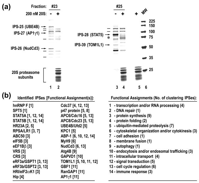
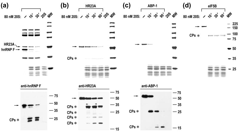
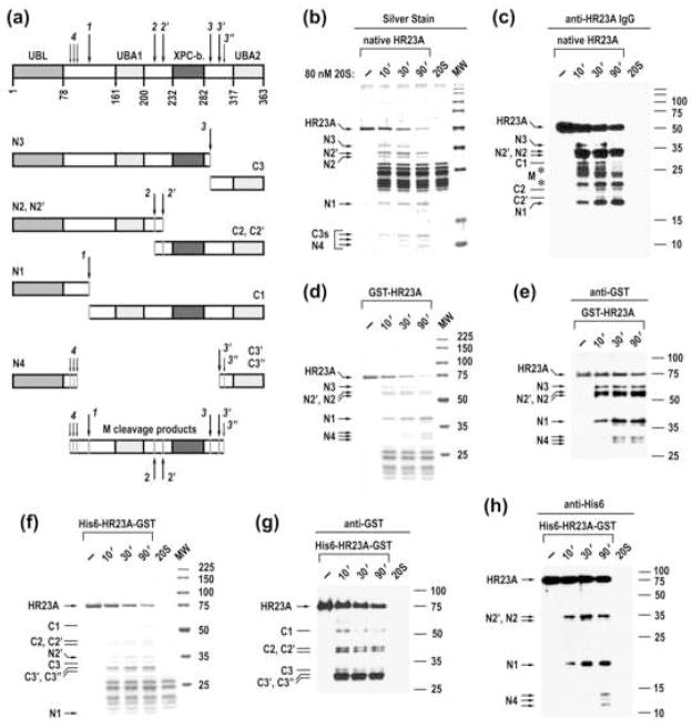
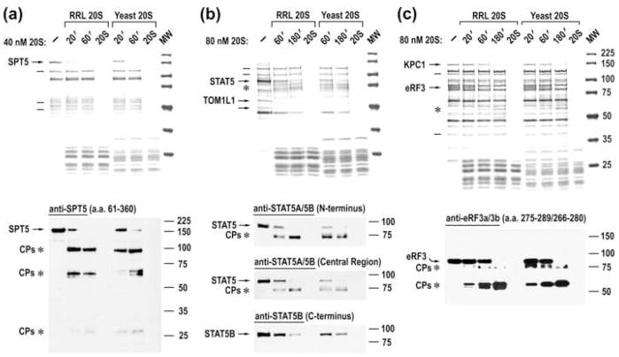
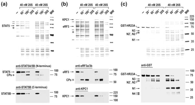
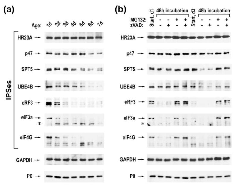
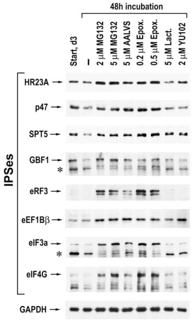
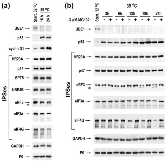
The critical role of the ubiquitin-26S proteasome system in regulation of protein homeostasis in eukaryotes is well established. In contrast, the impact of the ubiquitin-independent proteolytic activity of proteasomes is poorly understood. Through biochemical analysis of mammalian lysates, we find that the 20S proteasome, latent in peptide hydrolysis, specifically cleaves more than 20% of all cellular proteins. Thirty intrinsic proteasome substrates (IPSs) were identified and in vitro studies of their processing revealed that cleavage occurs at disordered regions, generating stable products encompassing structured domains. The mechanism of IPS recognition is remarkably well conserved in the eukaryotic kingdom, as mammalian and yeast 20S proteasomes exhibit the same target specificity. Further, 26S proteasomes specifically recognize and cleave IPSs at similar sites, independent of ubiquitination, suggesting that disordered regions likely constitute the universal structural signal for IPS proteolysis by proteasomes. Finally, we show that proteasomes contribute to physiological regulation of IPS levels in living cells and the inactivation of ubiquitin-activating enzyme E1 does not prevent IPS degradation. Collectively, these findings suggest a significant contribution of the ubiquitin-independent proteasome degradation pathway to the regulation of protein homeostasis in eukaryotes.


Figures

Fig. 1
Fig. 1
20S proteasome specifically cleaves multiple cellular proteins. (a) An example of analysis of cellular proteins for the presence of IPSes. Gel filtration elution fractions were incubated for 3 hours with or without 20S proteasomes and stained with Blue R, following SDS-PAGE. Protein species that were cleaved or remained intact are marked with arrows or lines to the left of the reactions. The position of new protein species that appeared after incubation with 20S proteasomes is marked with an asterisk. The last lane is a MW marker. (b) Summary of identified IPSes and their known functional engagements.
Fig. 2
Fig. 2
Analysis of IPS cleavage by 20S proteasome. Time courses of cleavage of partly purified HR23A and hnRNP F (a) and highly purified proteins (b–d). Reactions were assembled as indicated above the panels. Upper panels, Blue R stained SDS-PAGE; bottom panels, immunoblot analysis. The positions of intact IPSes (arrows) and cleavage products (CPs, asterisks) are marked to the left of the panels.
Fig. 3
Fig. 3
20S proteasome cleaves HR23A at internal disordered regions while sparing structured domains. (a) The model of HR23A cleavage. The upper lane schematically represents the structure of HR23A, the empty boxes denote flexible regions and the filled boxes correspond to the ubiquitin-like (UBL), ubiquitin associated (UBA), and XPC protein-binding (XPC-b) domains. Positions of cleavage sites (arrows with numbers) are tentative, based upon the mapping of cleavage products with specific antibodies and the mobility of the products. (b, c) Time course of cleavage of native rabbit HR23A protein. Reactions were assembled as indicated and analyzed by silver staining and western blotting with anti-serum to human HR23A protein. The assignment of cleavage products is based upon their mobility and time course of their generation. Cleavage products that likely comprise the middle portion of HR23A are marked with asterisks. (d, e) Time course of cleavage of GST-human HR23A recombinant protein. (f–h) Time course of cleavage of His6-human HR23A-GST. Reactions were stained with Blue R or probed with antibodies specific to terminal tags as indicated above the panels. The positions of C-terminal (Cx) and N-terminal (Nx) cleavage products are marked with lines and arrows, respectively, to the left of the panels.
Fig. 4
Fig. 4
Mammalian and yeast 20S proteasomes specifically recognize and cleave the same mammalian IPSes. (a–c) Blue R staining of SDS-PAGE of time courses of cleavage of partly purified rabbit IPSes with rabbit or yeast 20S proteasomes (upper panels) and immunoblot analysis of these reactions (bottom panels). The positions of IPSes (arrows), their cleavage products (asterisks), and some of the stable cellular proteins (lines) are marked to the left of the panels.
Fig. 5
Fig. 5
26S and 20S proteasomes specifically cleave the same IPSes. Blue R staining of SDS-PAGE of time courses of cleavage of partly purified native IPSes (a, b) and recombinant GST-HR23A (c) with 20S and 26S proteasomes (upper panels) and immunoblot analysis of these reactions (bottom panels).
Fig. 6
Fig. 6
Proteasomes differentially regulate levels of different IPSes. (a) Immunoblot analysis of LoVo lysates harvested at different days, using antibodies specific to different IPSes as well as to GAPDH and ribosomal protein P0. (b) Immunoblot analysis of LoVo lysates harvested at different days following treatment with 5 μM MG132 and 50 μM z-VAD-FMK as indicated above the panel. The positions of intact proteins and their cleavage products (asterisk) are indicated to the left of the panel.
Fig. 7
Fig. 7
The effect of different proteasome inhibitors on IPS levels. LoVo cells were treated as indicated above the panels and the lysates were analyzed by western blotting, using antibodies specific to the indicated proteins.
Fig. 8
Fig. 8
Proteasomes degrade many IPSes in a ubiquitin-independent manner. (a) ts85 cells were grown one day at the permissive temperature (32°C) and harvested immediately (start) or after 16 and 24 hours of further growth at the restrictive temperature (39°C). Cellular lysates were analyzed by western blotting, using antibodies specific to the indicated proteins. (b) Immunoblot analysis of ts85 cells grown at 32°C and harvested immediately and at different time points after further growth at 39°C in the absence or in the presence of 3 μM MG132.

References

    1. Ciechanover A. Intracellular protein degradation: from a vague idea thru the lysosome and the ubiquitin-proteasome system and onto human diseases and drug targeting. Hematology. 2006;2006:1–12. - PubMed
    1. Goldberg AL. Functions of the proteasome: from protein degradation and immune surveillance to cancer therapy. Biochem Soc Trans. 2007;35:12–17. - PubMed
    1. Wolf DH, Hilt W. The proteasome: a proteolytic nanomachine of cell regulation and waste disposal. Biochim Biophys Acta. 2004;1695:19–31. - PubMed
    1. Bajorek M, Glickman MH. Keepers at the final gates: regulatory complexes and gating of the proteasome channel. Cell Mol Life Sci. 2004;61:1579–1588. - PMC - PubMed
    1. DeMartino GN, Gillette TG. Proteasomes: machines for all reasons. Cell. 2007;129:659–662. - PubMed

Publication types