Review
. 2009 Sep 1;162(3):787-96.
doi: 10.1016/j.neuroscience.2008.12.042. Epub 2008 Dec 30.

The cerebellum and eye-blink conditioning: learning versus network performance hypotheses

Affiliations
Review

The cerebellum and eye-blink conditioning: learning versus network performance hypotheses

V Bracha et al. Neuroscience. .

Abstract

Classical conditioning of the eye-blink reflex in the rabbit is a form of motor learning that is uniquely dependent on the cerebellum. The cerebellar learning hypothesis proposes that plasticity subserving eye-blink conditioning occurs in the cerebellum. The major evidence for this hypothesis originated from studies based on a telecommunications network metaphor of eye-blink circuits. These experiments inactivated parts of cerebellum-related networks during the acquisition and expression of classically conditioned eye blinks in order to determine sites at which the plasticity occurred. However, recent evidence revealed that these manipulations could be explained by a network performance hypothesis which attributes learning deficits to a non-specific tonic dysfunction of eye-blink networks. Since eye-blink conditioning is mediated by a spontaneously active, recurrent neuronal network with strong tonic interactions, differentiating between the cerebellar learning hypothesis and the network performance hypothesis represents a major experimental challenge. A possible solution to this problem is offered by several promising new approaches that minimize the effects of experimental interventions on spontaneous neuronal activity. Results from these studies indicate that plastic changes underlying eye-blink conditioning are distributed across several cerebellar and extra-cerebellar regions. Specific input interactions that induce these plastic changes as well as their cellular mechanisms remain unresolved.


Figures

Fig. 1
Fig. 1
Schematic of the eyeblink conditioning paradigm. A. Rabbits are presented with a paired tone conditioned stimulus (CS) and airpuff unconditioned stimulus (US). Evoked eyeblinks are recorded with an infrared sensor. B. Idealized eyeblink records in naive and trained animals and the pulse diagram denoting the timing of stimuli. In the delay classical conditioning paradigm, the onset of the CS precedes the onset of the US and the stimuli co-terminate. Naive animals don’t respond to the CS, but the US evokes reliably the hard-wired trigeminal unconditioned blink (UR, top eyeblink trace). Over time, rabbits associate the CS with the US, and they learn to blink in anticipation of the upcoming aversive US. These associatively learned responses are called conditioned responses (CR, the second trace from the top).
Fig. 2
Fig. 2
A conceptual block diagram of the cerebellum-related circuitry involved in acquisition and expression of classically conditioned eyeblinks in the rabbit. This diagram is a highly simplified representation of relevant structures and connectivity. Information regarding the conditioned stimulus (CS) and unconditioned stimulus (US) information enters the network via auditory and sensory trigeminal systems. These inputs are supplied in parallel to the serially connected pontine nuclei, cerebellar cortex, cerebellar interposed nuclei (IN) and brainstem nuclei contributing to projections to eyeblink premotoneurons and supplying motor commands to them. Since all these sites (labeled with a star) receive CS and US information, they should be considered as putative sites of learning. Output of eyeblink premotoneurons supplies motor commands to eyeblink motoneurons. Backslashed circles denote nodes at which inactivation during training disrupts CR acquisition. Boxes with bold borders represent structures among which are in our view distributed plastic changes underlying eyeblink conditioning. BC – brachium conjunctivum; PM – nuclei containing eyeblink premotoneurons that include the red nucleus. The plus symbols mark excitatory glutamatergic inputs and minus signs label inhibitory GABA-ergic inputs.
Fig. 3
Fig. 3
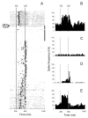
An example of the parallel effects of inferior olivary NBQX infusion on CR performance and on the activity of a task-modulated IN cell. This experiment consisted of 260 trials. After 40 baseline trials, NBQX was injected at the beginning of a 40-trial no-stimulation period. A, Raster plot of IN cell activity during this experiment. The experiment starts at the top with each row representing one trial, and each dot marking the occurrence of an action potential. The black square in each row corresponds to the onset of the eyeblink in that particular trial. Consequently, CRs have onset markers between lines denoting the CS and US onsets. Eyeblinks initiated past the US onset occur in trials in which the animal failed to produce the CR. Black squares at the ends of the 40 trials following the NBQX injection marker denote the no-stimulation waiting period that was inserted to allow for drug diffusion. Before the injection, this cell responded with excitation during the CS–US interval and with a combined excitatory/inhibitory response to the US. During the drug diffusion period, the firing rate of this cell’s activity precipitously declined. When stimulation was resumed, CRs were abolished immediately as evidenced by the blink onset marks on the right side of the US onset line. The baseline activity remained suppressed, and modulation during the CS–US interval was severely reduced whereas the relative excitatory modulation to the US became more distinct. The neuronal activity gradually recovered toward the end of the experiment in parallel with the recovery of behavioral CRs. B–E, Peri-stimulus histograms of the same IN unit constructed for 40 trials before the injection (B), for 40 post-injection drug diffusion trials when stimulation was paused (C), for 40 trials following the waiting period when stimulation was resumed (D), and for the last 40 trials from the remaining 140 trials of the experiment (E). Bin width for histograms in B–E is 20 ms. CS, onset of conditioned stimulus; US, onset of unconditioned stimulus. (Reprinted with permission from Zbarska et al., 2008)
Fig. 4
Fig. 4
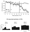
Effects of injecting the IN with the chloride channel blocker, picrotoxin (PTX), on the expression of CRs and on IN neuronal activity. Top panel: CR incidence in 15 injection experiments in which two injections of PTX were applied to the IN. The first injection (I1) had only a small effect on the frequency of CRs. A more extensive block of GABA-ergic neurotransmission with the second PTX injection (I2) gradually abolished CRs. Control injections of vehicle (aCSF) did not affect CR incidence. Bottom panel: population peri-stimulus histograms of 55 neurons recorded during PTX injections. Before injections, this population exhibited approximately a 25 Hz spontaneous firing rate and an excitatory response in the CS-US interval. The first PTX injection doubled the spontaneous discharge of IN neurons and reduced their CS-related modulation. Following the second injection, the spontaneous activity further increased, and the responses in the CS-US interval were almost completely attenuated. Two horizontal lines in each histogram denote tolerance limits used for detecting significant levels of neuronal modulation relative to mean baseline activity (Adapted from Aksenov et al., 2004).

Similar articles

Cited by

References

    1. Aksenov D, Serdyukova N, Irwin K, Bracha V. GABA neurotransmission in the cerebellar interposed nuclei: involvement in classically conditioned eyeblinks and neuronal activity. J Neurophysiol. 2004;91:719–727. - PubMed
    1. Aksenov D, Serdyukova N, Bloedel JR, Bracha V. 2004 Abstract Viewer/Itinerary Planner. Washington, DC: Society for Neuroscience; 2004. The role of tonic activity and phasic modulation of interposed cerebellar nuclear cells in the expression of conditioned eyeblinks. 136.6.
    1. Albus JS. A theory of cerebellar function. Math Biosci. 1971;10:25–61.
    1. Attwell PJ, Cooke SF, Yeo CH. Cerebellar function in consolidation of a motor memory. Neuron. 2002a;34:1011–1020. - PubMed
    1. Attwell PJ, Ivarsson M, Millar L, Yeo CH. Cerebellar mechanisms in eyeblink conditioning. Ann N Y Acad Sci. 2002b;978:79–92. - PubMed

Publication types