. 2009 Mar;40(3):390-400.
doi: 10.1016/j.mcn.2008.12.007. Epub 2008 Dec 31.

N-cadherin modulates voltage activated calcium influx via RhoA, p120-catenin, and myosin-actin interaction

Affiliations

N-cadherin modulates voltage activated calcium influx via RhoA, p120-catenin, and myosin-actin interaction

Glen S Marrs et al. Mol Cell Neurosci. 2009 Mar.

Abstract

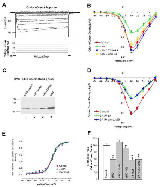
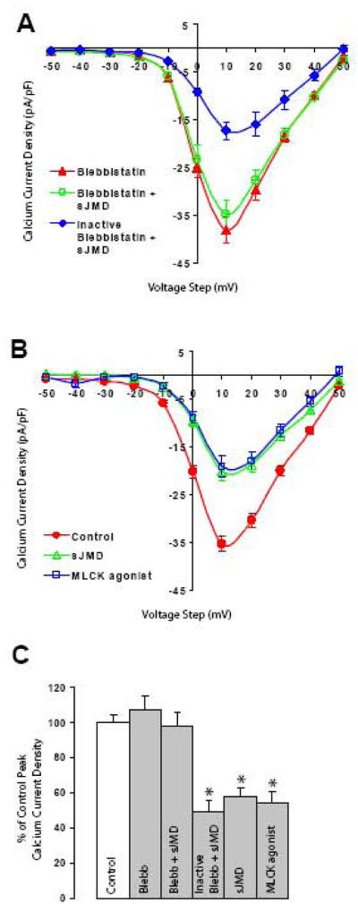
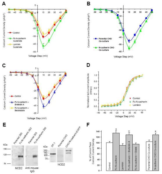
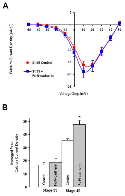
N-cadherin is a transmembrane adhesion receptor that contributes to neuronal development and synapse formation through homophilic interactions that provide structural-adhesive support to contacts between cell membranes. In addition, N-cadherin homotypic binding may initiate cell signaling that regulates neuronal physiology. In this study, we investigated signaling capabilities of N-cadherin that control voltage activated calcium influx. Using whole-cell voltage clamp recording of isolated inward calcium currents in freshly isolated chick ciliary ganglion neurons we show that the juxtamembrane region of N-cadherin cytoplasmic domain regulates high-threshold voltage activated calcium currents by interacting with p120-catenin and activating RhoA. This regulatory mechanism requires myosin interaction with actin. Furthermore, N-cadherin homophilic binding enhanced voltage activated calcium current amplitude in dissociated neurons that have already developed mature synaptic contacts in vivo. The increase in calcium current amplitude was not affected by brefeldin A suggesting that the effect is caused via direct channel modulation and not by increasing channel expression. In contrast, homotypic N-cadherin interaction failed to regulate calcium influx in freshly isolated immature neurons. However, RhoA inhibitors enhanced calcium current amplitude in these immature neurons, suggesting that the inhibitory effect of RhoA on calcium entry is regulated during neuronal development and synapse maturation. These results indicate that N-cadherin modulates voltage activated calcium entry by a mechanism that involves RhoA activity and its downstream effects on the cytoskeleton, and suggest that N-cadherin provides support for synaptic maturation and sustained synaptic activity by facilitating voltage activated calcium influx.


Figures

Figure 1
Figure 1. N-cadherin sJMD regulation of HVA Ca2+ current requires binding to p120-catenin and RhoA activation
A) Whole-cell voltage-clamp recordings of isolated inward Ca2+ currents from acutely dissociated ciliary ganglion neurons. Representative current response traces (upper panel) were elicited by 100 ms duration 10 mV steps in the voltage holding potential sequentially from −50mV to 50mV (lower panel). B) Current density plot shows averaged peak current reduction associated with infusion of N-cadherin sJMD as compared with control solution (control circles 35.4 ± 1.6, n=21; sJMD (1μM) open triangles 20.4 ± 1.8, n=12). Application of sJMD780AAA has no effect on current density (sJMD 780AAA (1μM) squares 38.4 ± 2.6, n=8). Simultaneous application of sJMD and C3 exotransferase substantially reverts peak current reduction associated with sJMD (sJMD (1μM) + C3 (0.5μM) open diamonds 32.5 ± 2.7, n=6). C) GST-tagged sJMD and GST-sJMD-780AAA were incubated with recombinant 6XHis-tagged p120-catenin (p120-228). Protein complexes were pulled down with glutathione-coated agarose beads, and analyzed by Western bolt with anti-His antibodies to detect p120-catenin. Lane 1, 5 μg of recombinant p120-228 runs at its predicted size of 89kDa. p120-catenin pulled down by recombinant GST (lane 2), sJMD-780AAA (lane 3), and sJMD (lane 4). Mutation of the p120-catenin binding site (sJMD-780AAA) substantially reduces p120-catenin binding to sJMD. D) Application of a dominant-active form of RhoA (DA-RhoA) reduces peak Ca2+ currents amplitude (DA-RhoA (0.4μM) open squares 19.8 ± 2.7, n=8). Simultaneous application of DA-RhoA and sJMD shows that their inhibitory effects are not additive (DA RhoA (0.4μM) + sJMD (1μM) diamonds 20.8 ± 2.5, n=10). E) Voltage dependence of activation obtained by plotting normalized tail current amplitude (I/Imax) against the voltage step. The continuous lines represent a Boltzmann fit of the data for control (circles), sJMD (open triangles), and DA RhoA (open squares). No difference in the activation profile was detected between treatments. Values are expressed as mean ± SE. F) Summary histogram showing averaged peak Ca2+ current density values for each condition tested in C and E expressed as a percentage of controls (100%). * Note significant decreases in HVA Ca2+ current influx for sJMD (p<0.001), DA RhoA (p<0.001), and DA RhoA + sJMD (p<0.001) associated conditions.
Figure 2
Figure 2. N-cadherin sJMD mediated Ca2+ current reduction requires actin-myosin interaction
A) Bath application of blebbistatin has no measurable effect on averaged peak Ca2+ currents (blebbistatin (50μM) triangles 38.1 ± 2.7, n=11). The presence of blebbistatin during pipette infusion of sJMD prevents current reduction (blebbistatin (50μM) + sJMD (1 μM) open circles 34.8 ± 3, n=10), indicating that sJMD signaling requires myosin interaction with actin. Photo-inactivated blebbistatin does not block sJMD reduction in Ca2+ current amplitude (inactive blebbistatin (50μM) + sJMD (1 μM) diamonds 17.3 ± 2, n=6). B) Infusion of an agonist peptide for myosin light chain kinase (MLCK) reduces averaged peak Ca2+ influx (control, circles 35.4 ± 1.6, n=21; sJMD (1μM) open triangles 20.4 ± 1.8, n=12; MLCK agonist (1μM) open squares 19.3 ± 2.1, n=4). Values are expressed as mean ± SE. C) Summary histogram of results for averaged peak Ca2+ current density presented in A and B expressed as a percentage of control values (100%). Blebbistatin (p=0.36) and blebbistatin + sJMD (p=0.85) conditions are comparable to control, while inactivated blebbistatin + sJMD (p<0.001) and MLCK agonist (p<0.001) conditions show significant reduction. * p<0.001
Figure 3
Figure 3. N-cadherin homophilic binding enhances HVA Ca2+ current amplitude
A) N-cadherin ectodomain C-terminally fused to an IgG Fc fragment (Fc-N-cadherin, 20μg/ml) was used as a substrate for plating dissociated St 40 ciliary neurons. Con A or laminin coated cover slips were used as controls. Ca2+ current density was analyzed 1-3h after plating (Con A, circles 35.4 ± 1.6, n=21; Fc-N-cadherin, open diamonds 47.7 ± 2.9, n=13; laminin, open squares 32 ± 2, n=8). B) Dissociated ciliary neurons were plated on CHO cells stably expressing full-length chicken N-cadherin C-terminally fused to EGFP and Ca2+ current amplitude was analyzed 6-8h after plating. Parental CHO cells devoid of N-cadherin expression (shown in E) were used as control (parental CHO, circles 45 ± 2.4, n=12; N-cadherin CHO, triangles 54.9 ± 2.6, n=13). C) Pre-incubation with brefeldin A (5mg/mL, 2hr) did not block Fc-N-cadherin induced peak Ca2+ current enhancement (control Con A, circles 35.4 ± 1.6, n=21; Fc-N-cadherin + brefeldin A, open triangles 45.5 ± 2.8, n=8). In contrast, bath application of blebbistatin blocked Fc-N-cadherin-mediated enhancement of averaged peak Ca2+ current (Fc-N-cadherin + blebbistatin (50μM) open squares 33.4 ± 1.8, n=6). D) Voltage dependence of activation obtained by plotting normalized tail current amplitude (I/Imax) against the voltage step. The continuous lines represent a Boltzmann fit of the data for control (circles), Fc-N-cadherin substrate (diamonds), and laminin (squares). E) Western blot analysis of recombinant Fc-N-cadherin used in A and CHO cell lines used for co-cultures in B. Left panel, HEK293 conditioned medium from parental and Fc-N-cadherin transfected cells immuno-blotted with anti-N-cadherin antibodies (left) or anti-mouse Fc fragment (right). Right panel, homogenates from parental or N-cadherin-EGFP transfected CHO cells immuno-blotted with anti-N-cadherin antibodies. Chicken fibroblast cell line (DF1 cells) homogenate was used as control (lane 1). Parental CHO cells are devoid of N-cadherin (lane 2). N-cadherin-EGFP runs at approximately 140kDa (lane 3), the additional band detected at ~240kDa may represent N-cadherin dimers. Values are expressed as mean ± SE. F) Summary histogram of the average peak Ca2+ current densities for the results presented in A, B, and C expressed as a percentage of their corresponding controls (100%). Left group, Fc-N-cadherin substrate vs control * p<0.01; brefeldin A vs control * p<0.01. Right group, N-cadherin expressing CHO cells vs parental CHO cells * p<0.05.
Figure 4
Figure 4. N-cadherin homophilic binding does not enhance HVA Ca2+ currents in St 35 neurons
A) Voltage clamp recordings from St 35 ciliary neurons plated on control Con A substrate or on Fc-N-cadherin substrate (Fc-N-cadherin). St 35 neurons show no substantial modulation of HVA Ca2+ current when plated on Fc-N-cadherin with respect to control (control circles 17 ± 1.5, n=6; Fc-N-cadherin substrate squares 18.9 ± 2.5, n=7, p=0.27). Values are expressed as mean ± SE. B) Summary histogram of the average peak Ca2+ current density recorded from St 35 and St 40 neurons plated on Con A or on an N-cadherin substrate (data from Fig 3A and 4A). * p<0.01
Figure 5
Figure 5. Development of ciliary neurons correlates with redistribution of ezrin and F-actin
Ciliary ganglia from St 35 and St 40 embryos were double stained with anti-N-cadherin (a, b) and antiezrin antibodies (c, d). Ezrin is localized to the membrane at St 35 and it becomes mostly cytosolic by St 40, suggesting a decrease in endogenous RhoA activity at St 40 as compared to St 35. g, h) Ciliary ganglion labeled with phalloidin-Alexa 488 to visualize the subcellular distribution of F-actin. Arrowheads in all panels point to the cell membrane. Scale bar in e for a, c, and e = 5μm; scale bar in f for b, d, and f = 5 μm, scale bar in g and h = 5 μm.
Figure 6
Figure 6. RhoA mediated inhibition of HVA Ca2+ currents is developmental regulated
A) Voltage clamp recording form St 35 ciliary neurons. Application of C3 exotransferase or p120-catenin (p120-228) protein results in enhancement (64% and 49% respectively) of averaged peak Ca2+ current amplitude as compared to control solution (control open circles 17 ± 1.5, n=6; C3 open diamonds 27.8 ± 2, n=6; p120-228 open triangles 25.4 ± 2.2, n=7). B) Application of C3 exotransferase or p120-catenin into St 40 ciliary neurons has no significant effect on peak Ca2+ current amplitude (control circles 35.4 ± 1.6, n=21; C3 (0.5μM) diamonds 31.1 ± 3.1, n=10; p120-288 (1μM) triangles 31.8 ± 1.9, n=6). Values are expressed as mean ± SE. C) Summary histograms of peak Ca2+ current densities for conditions reported in A and B expressed as a percentage of their respective controls (100%). At St 35 peak Ca2+ current density was significantly enhanced for both C3 (* p<0.01) and p120-catenin (* p<0.01). In contrast, no significant effects were observed on St 40 neurons with both RhoA inhibitors (C3: 12% reduction, p=0.16; p120-catenin 10% reduction, p=0.27).
Figure 7
Figure 7. Diagram showing the element involved in the regulation of HVA Ca2+ influx downstream of RhoA activation by N-cadherin JMD
Infusion of sJMD into freshly dissociate St 40 ciliary neurons suppresses the inhibitory effect of p120-catenin on RhoA activity, presumably by disrupting a molecular complex required to inhibit RhoA. Application of a mutated sJMD (sJMD780AAA) which does not bind p120-catenin has no effect on Ca2+ influx. The activation of RhoA through this pathway negatively influences HVA Ca2+ influx by a mechanism that requires myosin interaction with actin. Co-application of sJMD with C3-exotransferase or blebbistatin suppresses the inhibitory effect of the sJMD on Ca2+ influx, indicating that the effect of sJMD is mediated by RhoA activation. In addition, both, DA-RhoA or an MLCK agonist, suppress Ca2+ influx similarly as infusion of sJMD.

Similar articles

Cited by

References

    1. Anastasiadis PZ, Reynolds AB. The p120 catenin family: complex roles in adhesion, signaling and cancer. J Cell Sci. 2000;113(Pt 8):1319–1334. - PubMed
    1. Anastasiadis PZ, Moon SY, Thoreson MA, Mariner DJ, Crawford HC, Zheng Y, Reynolds AB. Inhibition of RhoA by p120 catenin. Nat Cell Biol. 2000;2:637–644. - PubMed
    1. Aono S, Nakagawa S, Reynolds AB, Takeichi M. p120(ctn) acts as an inhibitory regulator of cadherin function in colon carcinoma cells. J Cell Biol. 1999;145:551–562. - PMC - PubMed
    1. Arikkath J, Reichardt LF. Cadherins and catenins at synapses: roles in synaptogenesis and synaptic plasticity. Trends Neurosci. 2008 - PMC - PubMed
    1. Augustine GJ, Charlton MP. Calcium dependence of presynaptic calcium current and post-synaptic response at the squid giant synapse. J Physiol. 1986;381:619–640. - PMC - PubMed

Publication types

MeSH terms