

Biofunctionalized electrospun silk mats as a topical bioactive dressing for accelerated wound healing
- PMID: 19162575
- PMCID: PMC2810481
- DOI: 10.1016/j.actbio.2008.12.013
Biofunctionalized electrospun silk mats as a topical bioactive dressing for accelerated wound healing
Abstract
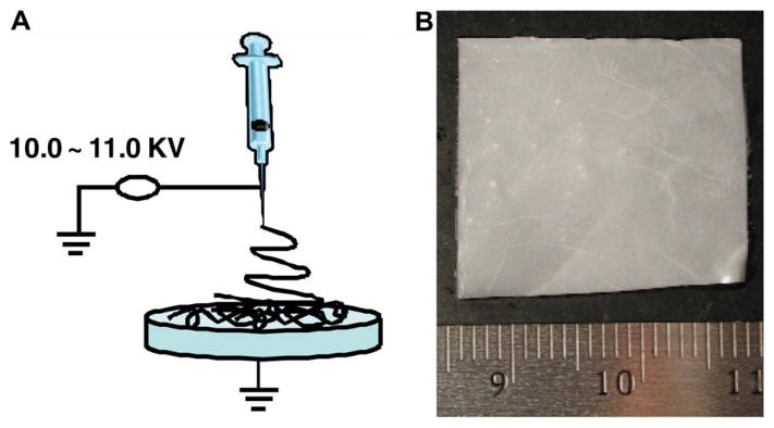
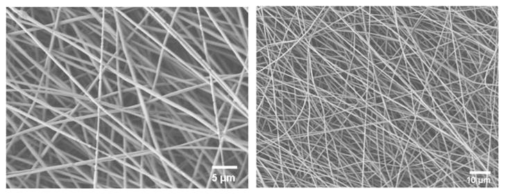
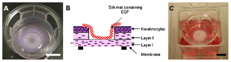
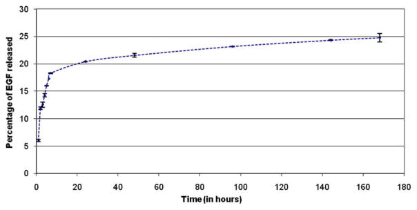
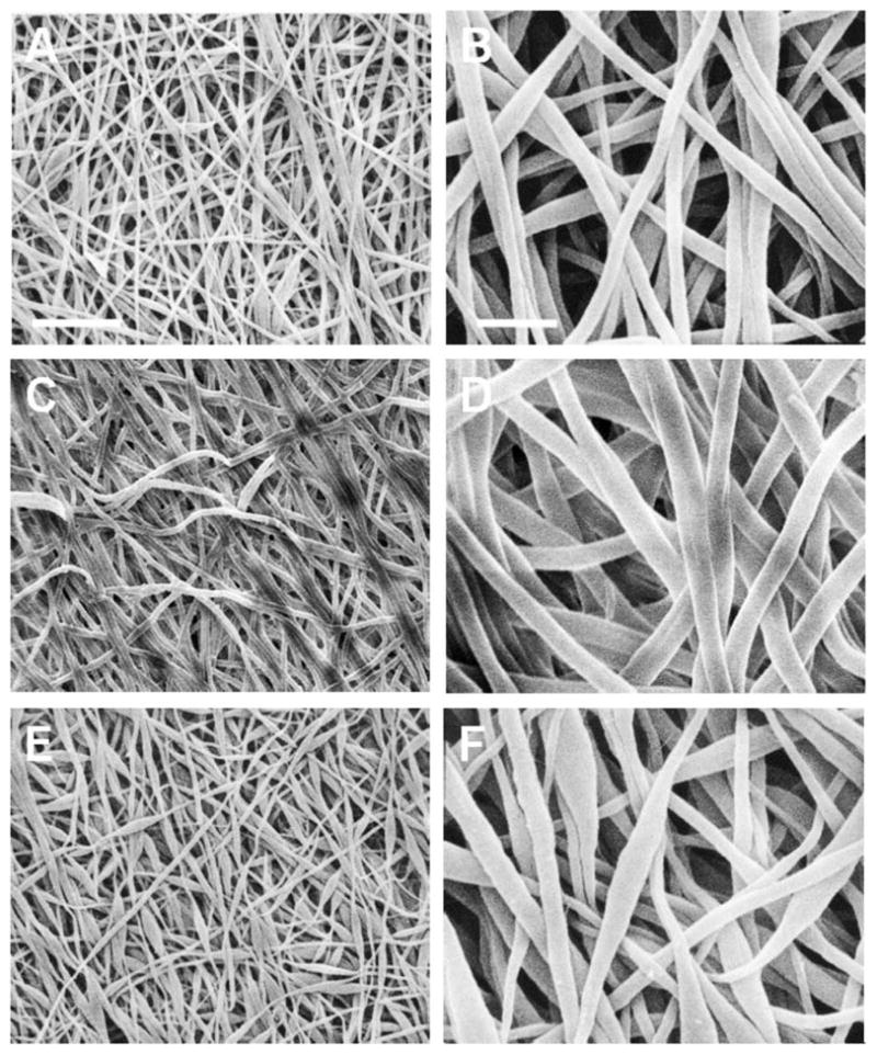
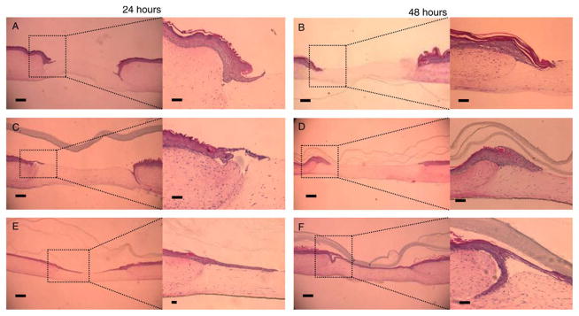
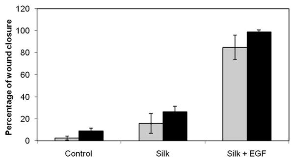
Materials able to deliver topically bioactive molecules represent a new generation of biomaterials. In this article, we describe the use of silk mats, made of electrospun nanoscale silk fibers containing epidermal growth factor (EGF), for the promotion of wound healing processes. In our experiments, we demonstrated that EGF is incorporated into the silk mats and slowly released in a time-dependent manner (25% EGF release in 170h). We tested these materials using a new model of wounded human skin-equivalents displaying the same structure as human skin and able to heal using the same molecular and cellular mechanisms found in vivo. This human three-dimensional model allows us to demonstrate that the biofunctionalized silk mats, when placed on the wounds as a dressing, aid the healing by increasing the time of wound closure by the epidermal tongue by 90%. The preservation of the structure of the mats during the healing period as demonstrated by electronic microscopy, the biological action of the dressing, as well as the biocompatibility of the silk demonstrate that this biomaterial is a new and very promising material for medical applications, especially for patients suffering from chronic wounds.
Figures
References
-
- Singer AJ, Clark RAF. Cutaneous wound healing. N Engl J Med. 1999;341:738–46. - PubMed
-
- Mustoe T. Understanding chronic wounds: a unifying hypothesis on their pathogenesis and implications for therapy. Am J Surg. 2004;187:65S–70S. - PubMed
-
- Winter GD. Epidermal regeneration in the domestic pig. In: Maibach HI, Rovee DT, editors. Epidermal wound healing. Chicago: Yearbook Medical; 1972. pp. 71–112.
-
- Sivamani RK, Garcia MS, Isseroff RR. Wound re-epithelialization: modulating keratinocyte migration in wound healing. Front Biosci. 2007;1:2849–68. - PubMed
-
- Dal Pra I, Chiarini A, Boschi A, Freddi G, Armato U. Novel dermoepidermal equivalents on silk fibroin-based formic acid-crosslinked three-dimensional nonwoven devices with prospective applications in human tissue engineering/regeneration/repair. Int J Mol Med. 2006;18:241–7. - PubMed
Publication types
MeSH terms
Substances
Grants and funding
LinkOut - more resources
Full Text Sources
