

Semi-automatic medical image segmentation with adaptive local statistics in Conditional Random Fields framework
- PMID: 19163362
- PMCID: PMC2882883
- DOI: 10.1109/IEMBS.2008.4649859
Semi-automatic medical image segmentation with adaptive local statistics in Conditional Random Fields framework
Abstract
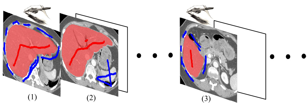
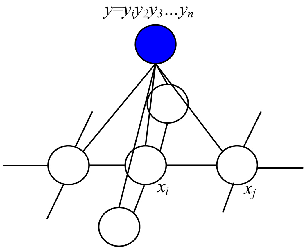
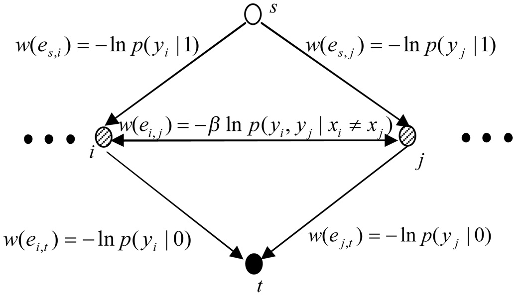
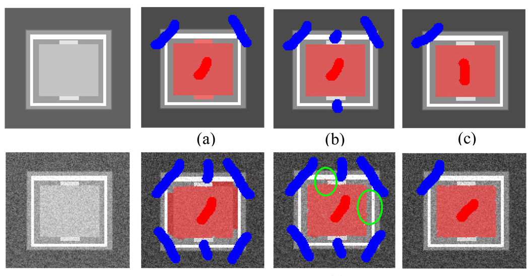
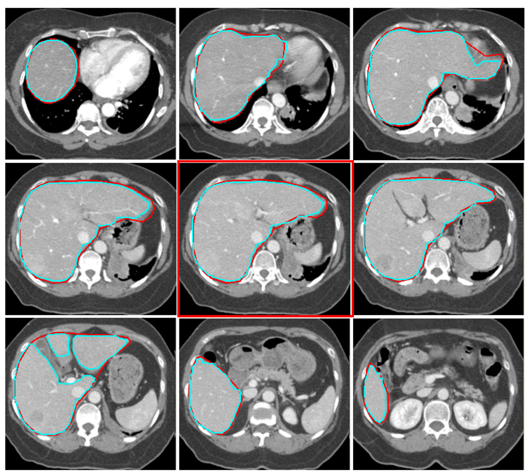
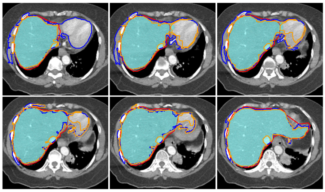
Planning radiotherapy and surgical procedures usually require onerous manual segmentation of anatomical structures from medical images. In this paper we present a semi-automatic and accurate segmentation method to dramatically reduce the time and effort required of expert users. This is accomplished by giving a user an intuitive graphical interface to indicate samples of target and non-target tissue by loosely drawing a few brush strokes on the image. We use these brush strokes to provide the statistical input for a Conditional Random Field (CRF) based segmentation. Since we extract purely statistical information from the user input, we eliminate the need of assumptions on boundary contrast previously used by many other methods, A new feature of our method is that the statistics on one image can be reused on related images without registration. To demonstrate this, we show that boundary statistics provided on a few 2D slices of volumetric medical data, can be propagated through the entire 3D stack of images without using the geometric correspondence between images. In addition, the image segmentation from the CRF can be formulated as a minimum s-t graph cut problem which has a solution that is both globally optimal and fast. The combination of a fast segmentation and minimal user input that is reusable, make this a powerful technique for the segmentation of medical images.
Figures

Similar articles
-
Interactive semiautomatic contour delineation using statistical conditional random fields framework.Med Phys. 2012 Jul;39(7):4547-58. doi: 10.1118/1.4728979. Med Phys. 2012. PMID: 22830786 Free PMC article.
-
Automatic segmentation of thoracic and pelvic CT images for radiotherapy planning using implicit anatomic knowledge and organ-specific segmentation strategies.Phys Med Biol. 2008 Mar 21;53(6):1751-71. doi: 10.1088/0031-9155/53/6/017. Epub 2008 Mar 7. Phys Med Biol. 2008. PMID: 18367801
-
Tumor segmentation with multi-modality image in Conditional Random Field framework with logistic regression models.Annu Int Conf IEEE Eng Med Biol Soc. 2014;2014:6450-4. doi: 10.1109/EMBC.2014.6945105. Annu Int Conf IEEE Eng Med Biol Soc. 2014. PMID: 25571473
-
Multi-level adaptive segmentation of multi-parameter MR brain images.Comput Med Imaging Graph. 2000 Mar-Apr;24(2):87-98. doi: 10.1016/s0895-6111(99)00042-7. Comput Med Imaging Graph. 2000. PMID: 10767588
-
A review of segmentation and deformable registration methods applied to adaptive cervical cancer radiation therapy treatment planning.Artif Intell Med. 2015 Jun;64(2):75-87. doi: 10.1016/j.artmed.2015.04.006. Epub 2015 May 16. Artif Intell Med. 2015. PMID: 26025124 Review.
Cited by
-
Semiautomatic tumor segmentation with multimodal images in a conditional random field framework.J Med Imaging (Bellingham). 2016 Apr;3(2):024503. doi: 10.1117/1.JMI.3.2.024503. Epub 2016 Jun 28. J Med Imaging (Bellingham). 2016. PMID: 27413768 Free PMC article.
-
Segmentation of Drosophila heart in optical coherence microscopy images using convolutional neural networks.J Biophotonics. 2018 Dec;11(12):e201800146. doi: 10.1002/jbio.201800146. Epub 2018 Aug 6. J Biophotonics. 2018. PMID: 29992766 Free PMC article.
-
Interactive semiautomatic contour delineation using statistical conditional random fields framework.Med Phys. 2012 Jul;39(7):4547-58. doi: 10.1118/1.4728979. Med Phys. 2012. PMID: 22830786 Free PMC article.
References
-
- Adams R, Bischof L. Seeded region growing. IEEE Trans. on Pattern Anal. Mach. Intell. 1994;16(6):641–647.
-
- Boykov Y, Jolly MP. Interactive graph cuts for optimal boundary & region segmentation of objects in N-D images. Proc. of Intl. Conf. Computer Vision. 2001;I:105–112.
-
- Boykov Y, Funka-Lea G. Graph cuts and efficient N-D image segmentation. Intl. Journal of Computer Vision. 2006;70:109–131.
-
- Boykov Y, Kolmogorov V. An experimental comparison of min-cut/ max-flow algorithms for energy minimization in vision. IEEE Trans. Pattern Anal. Mach. Intell. 2004;26(9):1124–1137. - PubMed
-
- Ford LR, Jr, Fulkerson DR. Maximal flow through a network. Canadian Journal of Math. 1956;8:399–404.
Publication types
MeSH terms
Grants and funding
LinkOut - more resources
Full Text Sources
Other Literature Sources
Medical