Impairment of postural control in rabbits with extensive spinal lesions
- PMID: 19164112
- PMCID: PMC2695648
- DOI: 10.1152/jn.00009.2008
Impairment of postural control in rabbits with extensive spinal lesions
Abstract
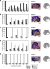
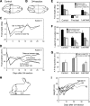
Our previous studies on rabbits demonstrated that the ventral spinal pathways are of primary importance for postural control in the hindquarters. After ventral hemisection, postural control did not recover, whereas after dorsal or lateral hemisection it did. The aim of this study was to examine postural capacity of rabbits after more extensive lesion (3/4 section of the spinal cord at T(12) level), that is, with only one ventral quadrant spared (VQ animals). They were tested before (control) and after lesion on the platform periodically tilted in the frontal plane. In control animals, tilts of the platform regularly elicited coordinated electromyographic (EMG) responses in the hindlimbs, which resulted in generation of postural corrections and in maintenance of balance. In VQ rabbits, the EMG responses appeared only in a part of tilt cycles, and they could be either correctly or incorrectly phased in relation to tilts. Because of a reduced value and incorrect phasing of EMG responses on both sides, this muscle activity did not cause postural corrective movements in the majority of rabbits, and the body swayed together with the platform. In these rabbits, the ability to perform postural corrections did not recover during the whole period of observation (< or =30 days). Low probability of correct EMG responses to tilts in most rabbits as well as an appearance of incorrect responses to tilts suggest that the spinal reflex chains, necessary for postural control, have not been specifically selected by a reduced supraspinal drive transmitted via a single ventral quadrant.
Figures


Similar articles
-
Effect of intrathecal administration of serotoninergic and noradrenergic drugs on postural performance in rabbits with spinal cord lesions.J Neurophysiol. 2008 Aug;100(2):723-32. doi: 10.1152/jn.90218.2008. Epub 2008 May 21. J Neurophysiol. 2008. PMID: 18497353 Free PMC article.
-
Impairment and recovery of postural control in rabbits with spinal cord lesions.J Neurophysiol. 2005 Dec;94(6):3677-90. doi: 10.1152/jn.00538.2005. Epub 2005 Jul 27. J Neurophysiol. 2005. PMID: 16049143
-
Facilitation of postural limb reflexes in spinal rabbits by serotonergic agonist administration, epidural electrical stimulation, and postural training.J Neurophysiol. 2011 Sep;106(3):1341-54. doi: 10.1152/jn.00115.2011. Epub 2011 Jun 8. J Neurophysiol. 2011. PMID: 21653706
-
Effect of acute lateral hemisection of the spinal cord on spinal neurons of postural networks.Neuroscience. 2016 Dec 17;339:235-253. doi: 10.1016/j.neuroscience.2016.09.043. Epub 2016 Oct 1. Neuroscience. 2016. PMID: 27702647 Free PMC article.
-
Postural control in the rabbit maintaining balance on the tilting platform.J Neurophysiol. 2003 Dec;90(6):3783-93. doi: 10.1152/jn.00590.2003. Epub 2003 Aug 20. J Neurophysiol. 2003. PMID: 12930819
Cited by
-
Changes in Activity of Spinal Postural Networks at Different Time Points After Spinalization.Front Cell Neurosci. 2019 Aug 21;13:387. doi: 10.3389/fncel.2019.00387. eCollection 2019. Front Cell Neurosci. 2019. PMID: 31496938 Free PMC article.
-
Effect of spinal cord injury on neural encoding of spontaneous postural perturbations in the hindlimb sensorimotor cortex.J Neurophysiol. 2021 Nov 1;126(5):1555-1567. doi: 10.1152/jn.00727.2020. Epub 2021 Aug 11. J Neurophysiol. 2021. PMID: 34379540 Free PMC article.
-
Role of CaMKIIa reticular neurons of caudal medulla in control of posture.bioRxiv [Preprint]. 2025 Mar 17:2025.03.17.643745. doi: 10.1101/2025.03.17.643745. bioRxiv. 2025. PMID: 40166225 Free PMC article. Preprint.
-
Common muscle synergies for balance and walking.Front Comput Neurosci. 2013 May 2;7:48. doi: 10.3389/fncom.2013.00048. eCollection 2013. Front Comput Neurosci. 2013. PMID: 23653605 Free PMC article.
-
Contribution of supraspinal systems to generation of automatic postural responses.Front Integr Neurosci. 2014 Oct 1;8:76. doi: 10.3389/fnint.2014.00076. eCollection 2014. Front Integr Neurosci. 2014. PMID: 25324741 Free PMC article. Review.
References
-
- Afelt Z Functional significance of ventral descending tracts of the spinal cord in the cat. Acta Neurobiol Exp 34: 393–407, 1974. - PubMed
-
- Akaike T, Westerman RA. Spinal segmental levels innervated by different types of vestibulo-spinal tract neurons in rabbit. Exp Brain Res 17: 443–446, 1973. - PubMed
-
- Barbeau H, Fung J, Leroux A, Ladouceur M. A review of the adaptability and recovery of locomotion after spinal cord injury. Progr Brain Res 137: 9–25, 2002. - PubMed
-
- Beloozerova IN, Zelenin PV, Popova LB, Orlovsky GN, Grillner S, Deliagina TG. Postural control in the rabbit maintaining balance on the tilting platform. J Neurophysiol 90: 3783–3793, 2003. - PubMed
-
- Blessing WW, Goodchild AK, Dampney RAL, Chalmers JP. Cell groups in the lower brain stem of the rabbit projecting to the spinal cord, with special reference to catecholamine-containing neurons. Brain Res 221: 35–55, 1981. - PubMed
Publication types
MeSH terms
Grants and funding
LinkOut - more resources
Full Text Sources
Medical

