. 2009 Apr;53(4):1598-609.
doi: 10.1128/AAC.01329-08. Epub 2009 Jan 21.

Genetic analysis of factors affecting susceptibility of Bacillus subtilis to daptomycin

Affiliations

Genetic analysis of factors affecting susceptibility of Bacillus subtilis to daptomycin

Anna-Barbara Hachmann et al. Antimicrob Agents Chemother. 2009 Apr.

Abstract

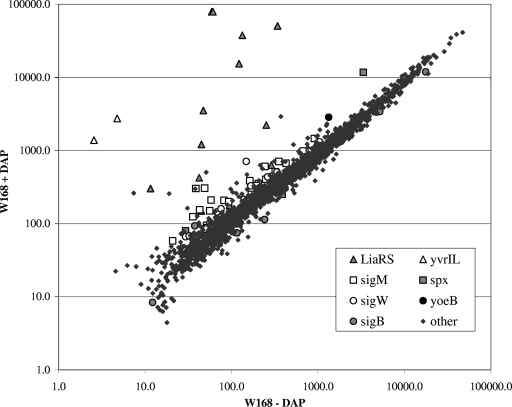
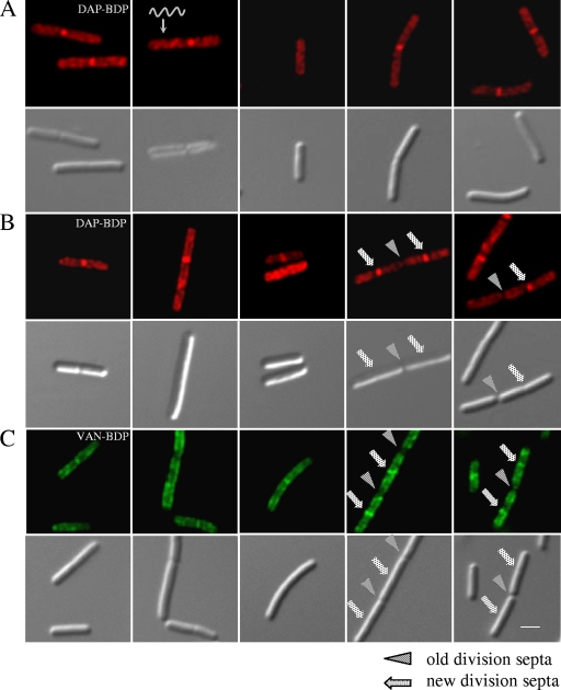
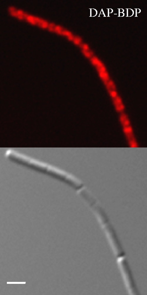
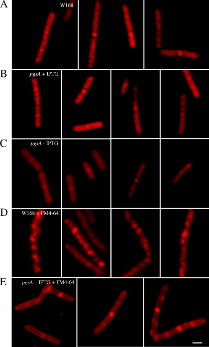
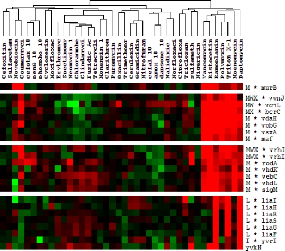
Daptomycin is the first of a new class of cyclic lipopeptide antibiotics used against multidrug-resistant, gram-positive pathogens. The proposed mechanism of action involves disruption of the functional integrity of the bacterial membrane in a Ca(2+)-dependent manner. We have used transcriptional profiling to demonstrate that treatment of Bacillus subtilis with daptomycin strongly induces the lia operon including the autoregulatory LiaRS two-component system (homologous to Staphylococcus aureus VraSR). The lia operon protects against daptomycin, and deletion of liaH, encoding a phage-shock protein A (PspA)-like protein, leads to threefold increased susceptibility. Since daptomycin interacts with the membrane, we tested mutants with altered membrane composition for effects on susceptibility. Deletion mutations of mprF (lacking lysyl-phosphatidylglycerol) or des (lipid desaturase) increased daptomycin susceptibility, whereas overexpression of MprF decreased susceptibility. Conversely, depletion of the cell for the anionic lipid phosphatidylglycerol led to increased resistance. Fluorescently labeled daptomycin localized to the septa and in a helical pattern around the cell envelope and was delocalized upon the depletion of phosphatidylglycerol. Together, these results indicate that the daptomycin-Ca(2+) complex interacts preferentially with regions enriched in anionic phospholipids and leads to membrane stresses that can be ameliorated by PspA family proteins.


Figures

FIG. 1.
FIG. 1.
Daptomycin stimulon in B. subtilis. The scatter plot represents the average expression levels of treated (+) versus untreated (−) cultures of B. subtilis W168 (daptomycin [DAP] at the MIC of 1 μg/ml; 20 min) from triplicate microarray analyses. The key lists highly expressed genes as grouped by their corresponding transcriptional regulators.
FIG. 2.
FIG. 2.
Daptomycin-BDP inserts preferentially at new division septa and in a spiral pattern. Fluorescent and DIC micrographs of B. subtilis stained with daptomycin-BDP (DAP-BDP) and vancomycin-BDP (VAN-BDP). (A) B. subtilis W168 treated with daptomycin-BDP at two times the MIC for 10 min (during exponential growth phase). (B) W168 treated with daptomycin-BDP at 10 times the MIC for 10 min. (C) W168 treated with equal amounts of vancomycin and vancomycin-BDP for 20 min. Panels A and B show a spiral localization of daptomycin-BDP and the preferential insertion at newly formed division septa, similar to that of vancomycin-BDP (C). The scale bar represents 2 μm.
FIG. 3.
FIG. 3.
Daptomycin-BDP (DAP-BDP) staining of stationary phase cells. Fluorescent and DIC micrographs of stationary growth phase culture of B. subtilis W168 treated with daptomycin-BDP at two times the MIC for 10 min. The staining shows a similar insertion pattern as that of exponential-growth phase cultures. The scale bar represents 2 μm.
FIG. 4.
FIG. 4.
Correlation between daptomycin-BDP staining and anionic phospholipid content and distribution. Fluorescent and DIC micrographs of wild-type W168 (A) and pgsA::pMutin grown in the presence (B) or absence (C) of 1 mM IPTG, stained with daptomycin-BDP at two times the MIC for 10 min. Daptomycin-BDP delocalizes when pgsA is not expressed from the IPTG-inducible promoter, suggesting preferential insertion of daptomycin in membrane lipid domains rich in anionic phosphatidylglycerol. W168 and pgsA::pMutin stained with the membrane lipid dye FM 4-64 in the absence of IPTG are shown in panels D and E, respectively. The scale bar represents 2 μm.
FIG. 5.
FIG. 5.
Cluster analysis of B. subtilis microarray studies with 40 different antimicrobial agents. The gene expression patterns of daptomycin-treated B. subtilis are most closely related to those of cells treated with the following cell membrane- and cell wall-active antibiotics: moenomycin, Triton X-114, polymyxin B, ramoplanin, ristocetin, and vancomycin. Cluster analysis was performed on whole genome data sets for each antibiotic (see Materials and Methods), and selected clusters enriched for daptomycin-induced genes are shown. Red indicates induction and green repression after treatment, whereas black corresponds to unchanged gene expression.

References

    1. Antelmann, H., S. Towe, D. Albrecht, and M. Hecker. 2007. The phosphorus source phytate changes the composition of the cell wall proteome in Bacillus subtilis. J. Proteome Res. 6:897-903. - PubMed
    1. Barák, I., K. Muchova, A. J. Wilkinson, P. J. O'Toole, and N. Pavlendova. 2008. Lipid spirals in Bacillus subtilis and their role in cell division. Mol. Microbiol. 68:1315-1327. - PMC - PubMed
    1. Bisicchia, P., D. Noone, E. Lioliou, A. Howell, S. Quigley, T. Jensen, H. Jarmer, and K. M. Devine. 2007. The essential YycFG two-component system controls cell wall metabolism in Bacillus subtilis. Mol. Microbiol. 65:180-200. - PubMed
    1. Butcher, B. G., and J. D. Helmann. 2006. Identification of Bacillus subtilis sigma-dependent genes that provide intrinsic resistance to antimicrobial compounds produced by bacilli. Mol. Microbiol. 60:765-782. - PubMed
    1. Campo, N., H. Tjalsma, G. Buist, D. Stepniak, M. Meijer, M. Veenhuis, M. Westermann, J. P. Muller, S. Bron, J. Kok, O. P. Kuipers, and J. D. Jongbloed. 2004. Subcellular sites for bacterial protein export. Mol. Microbiol. 53:1583-1599. - PubMed

Publication types