. 2009 May;80(5):1036-45.
doi: 10.1095/biolreprod.108.074658. Epub 2009 Jan 21.

Seminal fluid drives expansion of the CD4+CD25+ T regulatory cell pool and induces tolerance to paternal alloantigens in mice

Affiliations

Seminal fluid drives expansion of the CD4+CD25+ T regulatory cell pool and induces tolerance to paternal alloantigens in mice

Sarah A Robertson et al. Biol Reprod. 2009 May.

Abstract

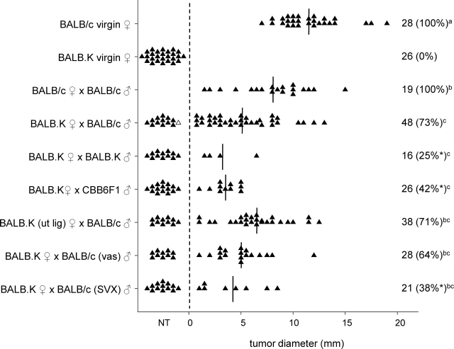
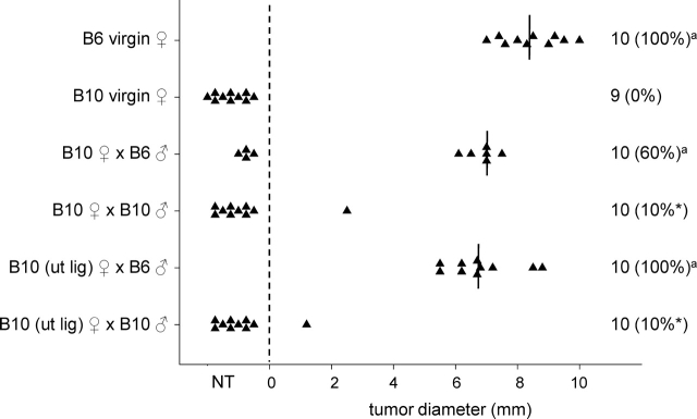
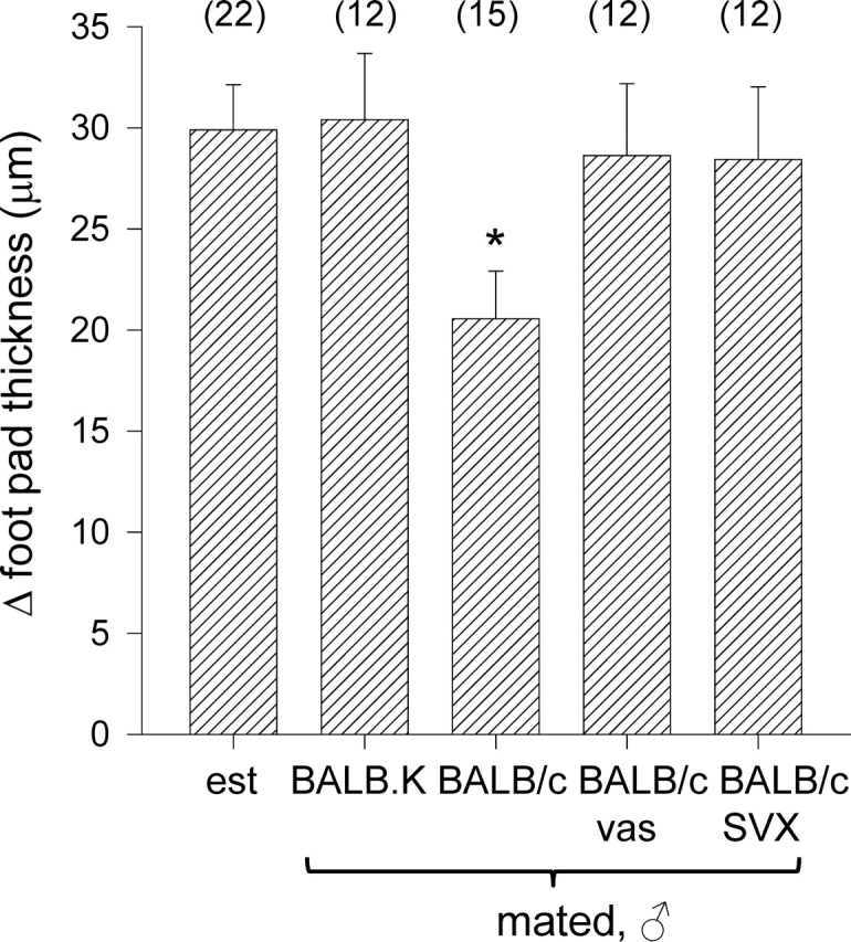
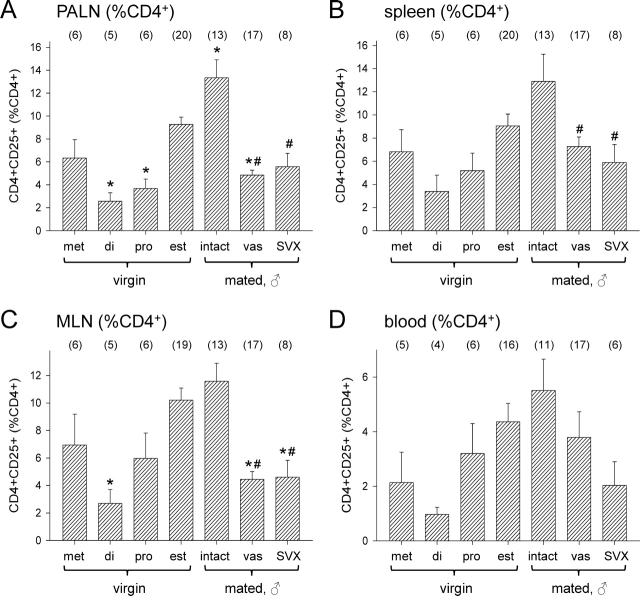
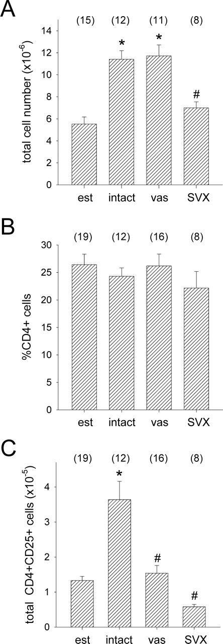
T regulatory (Treg) cells are implicated in maternal immune tolerance of the conceptus at implantation; however, the antigenic and regulatory signals controlling Treg cells in early pregnancy are undefined. To examine the role of male seminal fluid in tolerance induction, the effect of exposure to seminal fluid at mating on responsiveness to paternal alloantigens was examined using paternal tumor cell grafts and by delayed-type hypersensitivity (DTH) challenge on Day 3.5 postcoitum. Exposure to seminal fluid inhibited rejection of paternal tumor cells, independently of fertilization and embryo development, while seminal fluid from major histocompatability complex (MHC)-dissimilar males was less effective. Similarly, mating with intact males suppressed the DTH response to paternal alloantigens in an MHC-specific fashion. Excision of the seminal vesicle glands diminished the tolerance-inducing activity of seminal fluid. Mating with intact males caused an increase in CD4(+)CD25(+) cells expressing FOXP3 in the para-aortic lymph nodes draining the uterus, beyond the estrus-associated peak in cycling mice. The increase in CD4(+)CD25(+) cells was abrogated when males were vasectomized or seminal vesicles were excised. Collectively, these data provide evidence that exposure to seminal fluid at mating promotes a state of functional tolerance to paternal alloantigens that may facilitate maternal acceptance of the conceptus at implantation, and the effects of seminal fluid are likely to be mediated by expansion of the Treg cell pool. Both seminal plasma and sperm components of the seminal fluid are necessary to confer full tolerance and elicit the Treg cell response, potentially through provision of immune-deviating cytokines and antigens, respectively.


Figures

FIG. 1.
FIG. 1.
The effect of seminal fluid exposure at mating on rejection of paternal tumor cell grafts in BALB.K female mice. Intact or uterine-ligated BALB/c or BALB.K females, either virgin or Day 3.5 pc after mating with intact, vasectomized (vas) or SVX BALB/c, BALB.K or CBB6F1 males, were administered 1 × 10⁵ JR-5 tumor cells, and the occurrence and size of any resulting tumor was measured 13 days later. Symbols represent individual mice. The number of mice per group and the percent tumor growth (in parentheses) is shown. Vertical lines indicate mean values for mice with tumors. The effect of treatment on proportion of mice with tumors was analyzed by chi-square analysis (*P < 0.05, compared with BALB.K ♀ × BALB/c ♂ group). The effect of treatment on tumor size was analyzed by ANOVA and post hoc Sidak t-test. Different superscript letters indicate significant differences between groups (P < 0.05). NT, no tumor; ut lig, uterine ligated.
FIG. 2.
FIG. 2.
The effect of seminal fluid exposure at mating on rejection of paternal tumor cell grafts in B10 female mice. Intact or uterine-ligated B6 or B10 females, either virgin or Day 3.5 pc after mating with intact B6 or B10 males, were administered 5 × 10⁵ LLT cells, and the occurrence and size of any resulting tumor was measured 13 days later. Symbols represent individual mice. The number of mice per group and the percent tumor growth (in parentheses) is shown. Vertical lines indicate mean values for mice with tumors. The effect of treatment on proportion of mice with tumors was analyzed by chi-square analysis (*P < 0.05, compared with B10 ♀ × B6 ♂ group). aNo effect of treatment on tumor size was evident when data were analyzed by ANOVA and post hoc Sidak t-test. NT, no tumor; ut lig, uterine ligated.
FIG. 3.
FIG. 3.
The effect of seminal fluid exposure at mating on DTH responses in BALB.K female mice. BALB.K females were immunized by s.c. injection with BALB/c spleenocytes, left unmated (control; est, estrus) or mated 3–5 days later with either intact BALB.K males or intact, vasectomized (vas), or SVX BALB/c males, then challenged by s.c. injection to the footpad on Day 3.5 pc with BALB/c spleenocytes. Footpad thickness was measured 24 h later. Data are change in footpad thickness (mean ± SEM), calculated as the difference in thickness between spleenocyte-challenged and control footpads. The effect of treatment on DTH response was analyzed by ANOVA and post hoc Sidak t-test (*P < 0.05, compared with unmated virgin control group).
FIG. 4.
FIG. 4.
Flow cytometric analysis of CD4+CD25+ cells and FOXP3 expression in CD4+CD25+ cells and CD4+CD25 cells from para-aortic LN of virgin control B6 females (A, C, E, G), and Day 3.5 pc B6 females after mating with intact BALB/c males (B, D, F, H). Data shown are representative of n = 5–6 mice per group. The proportion of CD4+CD25+ cells as a percentage of CD4+ cells is shown in the top left corner of dot plots (AD). Histograms show the expression of FOXP3 in CD4+CD25+ cells (E and F) and in CD4+CD25 cells (G and H), with the percentage of FOXP3+ cells within each population shown in the top left corner. APC, allophycocyanin; est, estrus.
FIG. 5.
FIG. 5.
The effect of stage of estrous cycle and exposure to seminal vesicle fluid on CD4+CD25+ cell populations in female B6 mice. Para-aortic LN (PALN) (A), spleen (B), mesenteric LN (MLN) (C), and peripheral blood (D) cells were recovered at defined stages of the estrous cycle (di, diestrus; est, estrus; met, metestrus; pro, proestrus) or from Day 3.5 pc females after mating with intact, vasectomized (vas), or SVX BALB/c males, and the proportion of CD4+CD25+ cells as a percentage of CD4+ cells was evaluated by flow cytometry. Data are mean ± SEM, with the number of mice in each group shown in parentheses. The effect of treatment was analyzed by ANOVA and post hoc Sidak t-test (*P < 0.05 compared with estrus control group; #P < 0.05 compared with intact mated group).
FIG. 6.
FIG. 6.
The effect of exposure to seminal fluid on absolute total cell numbers (A), percent CD4+ cells (B), and absolute CD4+CD25+ cells (C) in para-aortic LN of female B6 mice. Cells were recovered from unmated control mice at estrus (est), or from Day 3.5 pc females after mating with intact, vasectomized (vas) or SVX BALB/c males, and absolute numbers were calculated from the percent CD4+CD25+ data shown in Figure 5A. Data are mean ± SEM, with the number of mice in each group shown in parentheses. The effect of treatment was analyzed by ANOVA and post hoc Sidak t-test (*P < 0.01, compared with estrus control group; #P < 0.01 compared with intact mated group).

Similar articles

Cited by

References

    1. Trowsdale J, Betz AG.Mother's little helpers: mechanisms of maternal-fetal tolerance. Nat Immunol 2006; 7: 241–246. - PubMed
    1. Guleria I, Sayegh MH.Maternal acceptance of the fetus: true human tolerance. J Immunol 2007; 178: 3345–3351. - PubMed
    1. Tafuri A, Alferink J, Moller P, Hammerling GJ, Arnold B.T cell awareness of paternal alloantigens during pregnancy. Science 1995; 270: 630–633. - PubMed
    1. Jiang SP, Vacchio MS.Multiple mechanisms of peripheral T cell tolerance to the fetal “allograft.” J Immunol 1998; 160: 3086–3090. - PubMed
    1. Zhou M, Mellor AL.Expanded cohorts of maternal CD8+ T-cells specific for paternal MHC class I accumulate during pregnancy. J Reprod Immunol 1998; 40: 47–62. - PubMed

Publication types