. 2009 Mar 27;284(13):8930-9.
doi: 10.1074/jbc.M809491200. Epub 2009 Jan 21.

A novel role for embigin to promote sprouting of motor nerve terminals at the neuromuscular junction

Affiliations

A novel role for embigin to promote sprouting of motor nerve terminals at the neuromuscular junction

Enzo Lain et al. J Biol Chem. .

Abstract

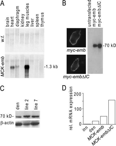
Adult skeletal muscle accepts ectopic innervation by foreign motor axons only after section of its own nerve, suggesting that the formation of new neuromuscular junctions is promoted by muscle denervation. With the aim to identify new proteins involved in neuromuscular junction formation we performed an mRNA differential display on innervated versus denervated adult rat muscles. We identified transcripts encoding embigin, a transmembrane protein of the immunoglobulin superfamily (IgSF) class of cell adhesion molecules to be strongly regulated by the state of innervation. In innervated muscle it is preferentially localized to neuromuscular junctions. Forced overexpression in innervated muscle of a full-length embigin transgene, but not of an embigin fragment lacking the intracellular domain, promotes nerve terminal sprouting and the formation of additional acetylcholine receptor clusters at synaptic sites without affecting terminal Schwann cell number or morphology, and it delays the retraction of terminal sprouts following re-innervation of denervated endplates. Conversely, knockdown of embigin by RNA interference in wild-type muscle accelerates terminal sprout retraction, both by itself and synergistically with deletion of neural cell adhesion molecule. These findings indicate that embigin enhances neural cell adhesion molecule-dependent neuromuscular adhesion and thereby modulates neuromuscular junction formation and plasticity.


Figures

FIGURE 1.
FIGURE 1.
Regulation of embigin expression in adult rodent muscle by electrical muscle activity. A, autoradiography of the initial mRNA differential display experiment. Three different samples of either innervated or denervated rat soleus muscles were used for PCR amplifications and analyzed on polyacrylamide gels. The differentially regulated PCR amplification product of 630 bp corresponds to embigin mRNA. B, denervation-induced transcription of embigin analyzed by Northern blot. Expression of the ribosomal protein L8 (RL8) was assessed as internal standard. C and D, transcript levels of embigin (white columns) and AChRd (black columns) were measured in rat (C) and in mouse (D) soleus muscles by quantitative RT-PCR on innervated (inn), denervated (d), and denervated/stimulated (den/stim) muscles at various time points after denervation. Expression levels were normalized to the expression of RL8. Innervated samples were arbitrarily set to 1, and -fold inductions ± S.E. are shown (n = 3-5). Open triangles indicate the percentage of ectopically innervated surface muscle fibers as a function of time after cutting the soleus nerve (data from Ref. 32). E, time course of embigin protein expression by Western blot following denervation in mouse leg muscle. Numbers give days after denervation. β-Tubulin was used as loading control. F, embigin mRNA relative to RL8 mRNA expression levels in neonatal (P0) compared with innervated and denervated adult rat (white columns) and mouse (black columns) leg muscles, analyzed by quantitative RT-PCR (n = 4).
FIGURE 2.
FIGURE 2.
Embigin is localized to the neuromuscular junction in neonatal (P0; A and B) and in adult (P126; C and D) soleus muscles. Embigin immunoreactivity (B and D) and synaptic AChR clusters resolved by α-Bgt staining (A and C) are shown in cross-sections of rat leg muscle. Incubation in primary antibody in the presence of immunizing peptide abolished synapse-specific staining (not shown). Bar:10 μm.
FIGURE 3.
FIGURE 3.
Embigin expression in MCK-emb transgenic mouse lines. A, Northern blot analysis in various organs of adult wild-type and MCK-emb mice shows strong embigin mRNA expression in the leg muscles of transgenic animals (line 3). B, protein products of transgenes used for setting up transgenic lines are directed to the cell membranes of HEK293 cells. The full-length and truncated embigin expression constructs tagged in c-Myc in their N termini were transfected into HEK293 cells. c-Myc-immunoreactivities in non-permeabilized HEK293 cells are shown. Western blot analysis confirms the specificity of the antibody for the c-Myc-tagged forms of embigin. The difference in calculated molecular weight between full-length (330 amino acids) and truncated embigin (290 amino acids) is not resolved on this blot because the deleted intracellular domain (calculated to be 4.5 kDa) is only 5% of the molecular mass of the glycolyated full-length protein with an molecular mass of 70 kDa (hence also called gp70), taking also the molecular weight of the Myc tags into account. C, Western blot analysis of embigin in mouse soleus muscles, using a rabbit polyclonal antibody. Two transgenic mouse lines (2 and 7) were tested, and expression levels were compared with a denervated wt sample. β-Actin was used as loading control. D, relative mRNA expression of embigin in denervated wild types, MCK-emb (line 2) and MCK-embDIC transgenic mice, measured by qRT-PCR. The value in innervated samples was set to 1.
FIGURE 4.
FIGURE 4.
Overexpression of embigin transgene in innervated soleus muscle induces nerve terminal sprouting that is dependent on the presence of embigin intracellular domain. A and B, in embigin full-length mutant animals, motoneurons (green) grow beyond the AChR clusters (red) stained with α-bungarotoxin-Alexa594. Sprouts are marked by arrows. The presence of small AChR clusters can be observed at the end of some sprouts (A, arrow). C, in comparison to wt littermates (left bar), the percentage of synapses exhibiting terminal sprouts is significantly increased in mice overexpressing full-length (center bar) but not truncated embigin (right bar). Only synapses with sprouts not apposed to AChR cluster such as in B were counted. Results are expressed as mean ± S.E. (n = 3-5; 21-78 analyzed synapses/animal; **, p < 0.01; line 2). Bar:10 μm.
FIGURE 5.
FIGURE 5.
Number of AChR clusters per synapse is increased in MCK-embigin soleus muscles. A, examples of endplates composed of different numbers of AChR clusters (given below the micrographs). B, in mutant mice (n = 10), the number of clusters per endplate was significantly increased in comparison to wt littermates (n = 9). An increase was observed also in MCK-MuSK mice (n = 6). Results are represented as mean ± S.E. (77-167 synapses analyzed/animal; **, p < 0.01; *, p < 0.05). Bar:10 μm.
FIGURE 6.
FIGURE 6.
The retraction of terminal sprouts following re-innervation is delayed in MCK-Emb mice (line 2). When endplates were re-innervated following nerve crush, sprouts grew beyond synaptic AChR clusters marked by staining with α-bungarotoxin-Alexa594 and then retracted (for illustration, see Fig. 7C). Retraction was delayed in MCK-emb mice. Bars show percentage of synapses 4 and 8 weeks after nerve crush. Results are expressed as mean ± S.E. (4 weeks: n = 6-7; 30 synapses analyzed/animal; **, p < 0.01; 8 weeks: mean from n = 2 animals is shown).
FIGURE 7.
FIGURE 7.
Inhibition of embigin expression by RNAi accelerates retraction of terminal sprouts at endplates re-innervated after nerve crush. A, test of efficiency to knockdown embigin expression by three different constructs encoding nuclear localisation signal-GFP and shRNA directed against embigin mRNA. HEK 293 cells were transfected with cDNA constructs encoding embigin alone or combined with one of three different constructs expressing shRNA (RNAi1, RNAi2, and RNAi3) designed to knock down embigin expression, and expression levels of embigin protein were analyzed by Western blot. Note that the intensity of the embigin band (70 kb) was strongly reduced after co-transfection with all three knockdown constructs. No reduction was seen after cotransfection of the same vector expressing shRNA against (unrelated) N-cadherin (RNAi N-cad). B, electroporation of the RNAi1 construct into mouse soleus muscles strongly reduces up-regulation or embigin protein by denervation. Den: denervated samples. β-Actin was used as loading control. C, example of terminal sprout (arrowhead) at re-innervated endplate 4 weeks after nerve crush. Axons were stained for neurofilaments (red) and synaptic AChRs with α-bungarotoxin-Alexa647 (blue), respectively. In electroporated muscles sprouts were counted only in fibers that had been transfected with knockdown constructs. They were marked by their expression of GFP (green). D, retraction of terminal sprouts formed at re-innervated endplates is accelerated by knockdown of embigin expression. The percentage of endplates exhibiting sprouts in the injected wild-type muscles (shRNA1-WT) was significantly lower than in the contralateral muscles (WT), which were taken as controls (n = 8; total of 163-191 synapses analyzed; **, p < 0.01). A further reduction in the number of endplates exhibiting sprouts was measured in transfected fibers of NCAM-deficient animals (shRNA1-NCAM-/-) in comparison to shRNA1-WT fibers (n = 8, 150-196 synapses; *, p < 0.05). No significant difference in the sprouting response between WT and NCAM-/- samples was resolved. Results are expressed as mean ± S.E. Bar:20 μm.
FIGURE 8.
FIGURE 8.
Schwann cell processes extending beyond the synaptic AChR cluster are present in equal measure in the soleus muscle of MCK-emb mice and wt littermates. A, Schwann cell processes marked by arrows. Bar, 10 μm. B, bars show percentage of synapses with sprouts (mean ± S.E.; n = 4; 143-145 synapses analyzed).

References

    1. Kim, N., and Burden, S. J. (2008) Nat. Neurosci. 11 19-27 - PMC - PubMed
    1. Bezakova, G., and Ruegg, M. A. (2003) Nat. Rev. Mol. Cell. Biol. 4 295-308 - PubMed
    1. Lømo, T. (2003) J. Neurocytol. 32 835-848 - PubMed
    1. Bowen, D. C., Park, J. S., Bodine, S., Stark, J. L., Valenzuela, D. M., Stitt, T. N., Yancopoulos, G. D., Lindsay, R. M., Glass, D. J., and DiStefano, P. S. (1998) Dev. Biol. 199 309-319 - PubMed
    1. Meier, T., Marangi, P. A., Moll, J., Hauser, D. M., Brenner, H. R., and Ruegg, M. A. (1998) Eur. J. Neurosci. 10 3141-3152 - PubMed

Publication types

MeSH terms