. 2009 Mar 20;284(12):7853-65.
doi: 10.1074/jbc.M808824200. Epub 2009 Jan 21.

Neutralization of interleukin-18 ameliorates ischemia/reperfusion-induced myocardial injury

Affiliations

Neutralization of interleukin-18 ameliorates ischemia/reperfusion-induced myocardial injury

Kaliyamurthi Venkatachalam et al. J Biol Chem. .

Abstract

Ischemia/reperfusion (I/R) injury is characterized by the induction of oxidative stress and proinflammatory cytokine expression. Recently demonstrating that oxidative stress and TNF-alpha each stimulate interleukin (IL)-18 expression in cardiomyocytes, we hypothesized that I/R also induces IL-18 expression and thus exacerbates inflammation and tissue damage. Neutralization of IL-18 signaling should therefore diminish tissue injury following I/R. I/R studies were performed using a chronically instrumented closed chest mouse model. Male C57BL/6 mice underwent 30 min of ischemia by LAD coronary artery ligation followed by various periods of reperfusion. Sham-operated or ischemia-only mice served as controls. A subset of animals was treated with IL-18-neutralizing antibodies 1 h prior to LAD ligation. Ischemic LV tissue was used for analysis. Our results demonstrate that, compared with sham operation and ischemia alone, I/R significantly increased (i) oxidative stress (increased MDA/4-HNE levels), (ii) neutrophil infiltration (increased MPO activity), (iii) NF-kappaB DNA binding activity (p50, p65), and (iv) increased expression of IL-18Rbeta, but not IL-18Ralpha or IL-18BP transcripts. Administration of IL-18-neutralizing antibodies significantly reduced I/R injury measured by reduced infarct size (versus control IgG). In isolated adult mouse cardiomyocytes, simulated ischemia/reperfusion enhanced oxidative stress and biologically active IL-18 expression via IKK-dependent NF-kappaB activation. These results indicate that IL-18 plays a critical role in I/R injury and thus represents a promising therapeutic target.


Figures

FIGURE 1.
FIGURE 1.
Reperfusion following 30 min of ischemia stimulates free radical generation and neutrophil infiltration. Male C57Bl/6 mice underwent 30 min of LAD coronary artery ligation, followed by reperfusion for various periods of time. A, details of the I/R protocol are shown. B, I/R induced free radical generation. Free radical generation was analyzed at 2 h of reperfusion by measuring the lipid peroxidation products MDA and 4-HNE in postischemic reperfused LV as described under “Experimental Procedures.” Ischemia alone and sham-operated animals served as controls. *, p < 0.001 (versus sham-operated; n = 4/group). C, ischemia/reperfusion induced neutrophil infiltration. MPO, a biochemical marker for neutrophil infiltration into tissues, was measured in postischemic reperfused myocardium using a colorimetric assay, as described under “Experimental Procedures.” *, p < 0.001 (versus sham and 30 min of ischemia alone).
FIGURE 2.
FIGURE 2.
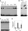
Ischemia/reperfusion activates NF-κB in postischemic myocardium. A, I/R induced NF-κB DNA-binding activity. Following 30 min of ischemia/2 h of reperfusion, nuclear proteins were extracted from postischemic reperfused LV tissue and analyzed for NF-κB DNA binding activity by EMSA as described under “Experimental Procedures.” Sham-operated and ischemia-alone groups served as controls (n = 4/group). Control studies determining the specificity of NF-κB oligonucleotide was shown in the completion experiments (lanes 1–4). Lane 1, protein extract from 30-min ischemia/2-h reperfusion myocardium was preincubated with a 75-fold molar excess of unlabeled double-stranded consensus NF-κB oligonucleotide, followed by the addition of labeled κB-specific probe. Lane 2, competition with cold mutant NF-κB oligonucleotide. Protein extract from 30-min ischemia/2-h reperfusion myocardium was preincubated with a 75-fold molar excess of unlabeled double-stranded mutant NF-κB oligonucleotide, followed by the addition of labeled κB-specific probe. Lane 3, protein extract from 30-min ischemia/2-h reperfusion myocardium was preincubated with a 75-fold molar excess of unlabeled double-stranded consensus Oct1 oligonucleotide, followed by the addition of labeled κB-specific probe. Lane 4, no nuclear protein, but the sample contains [γ-32P]ATP-labeled κB-specific probe. Lane 5, protein extract from 30-min ischemia/2-h reperfusion myocardium was incubated with 32P-labeled mutant NF-κB oligonucleotide. Lanes 6–9, protein extract from LV tissue from Sham-operated animals + κB-specific probe. Lanes 10–13, protein extract from ischemic LV tissue from 30 min of ischemia + κB-specific probe. Lanes 14–17, protein extract from LV tissue from postischemic reperfused LV tissue + κB-specific probe. Solid arrow, NF-κB-specific DNA-protein complexes. Solid circle, free probe. B, I/R-induced NF-κB activation was composed of p50 and p65. The nuclear extracts used in lane 14 in A were preincubated with p50 or p65 antibodies for 40 min prior to incubation with κB-specific probe followed by EMSA (n = 3). Normal IgG served as a control (Control IgG). Solid arrow, NF-κB-specific binding region. Open arrow, supershifted bands. Solid circle, free probe. C, I/R had no effect on basal Oct1 DNA binding activity. Nuclear protein extracts described in A were analyzed for Oct1 DNA binding by EMSA (n = 3). Solid arrow, Oct1-specific DNA-protein complexes. Solid circle, free probe. D, I/R-induced NF-κB activation was quantified by ELISA. Nuclear extracts described in A were analyzed for NF-κB activation and subunit composition by ELISA, as described under “Experimental Procedures” (n = 4/group, p < at least 0.001 versus corresponding Sham or ischemia (I) alone).
FIGURE 3.
FIGURE 3.
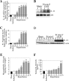
Ischemia/reperfusion induces IL-18 expression in serum and postischemic myocardium. A, I/R induced IL-18 mRNA expression in a time-dependent manner. IL-18 mRNA in total RNA extracts was quantified by RT-qPCR. β-actin served as an internal control. *, p < 0.05; **, p < 0.001 versus sham and ischemia (I) alone (n = 4/group). B, I/R-induced IL-18 mRNA expression at 2 h of reperfusion was confirmed by Northern blot analysis. Each lane contains 20 μg of total RNA from an individual animal. 28 S rRNA served as an internal control and shows similar levels of RNA loading in each lane. C, I/R induced IL-18 protein expression in a time-dependent manner. IL-18 protein levels were quantified by immunoblotting. *, p < 0.01; **, p < 0.001 versus sham and ischemia (I) alone (n = 4/group). Representative immunoblotting at 2 h of reperfusion is shown in D. Each lane contains 30 μg of protein extract from an individual animal. β-Actin served as an internal control and shows similar levels of protein loading in each well. E, IL-18 levels in myocardial protein extracts were quantified by ELISA. *, p < 0.05; **, p < at least 0.01 versus Sham and ischemia (I) alone (n = 4/group). F, I/R enhanced systemic IL-18 levels in a delayed manner. Serum levels of IL-18 at the indicated time periods were quantified by ELISA. *, p < 0.05; **, p < 0.01 versus sham and ischemia (I) alone (n = 4/group).
FIGURE 4.
FIGURE 4.
I/R induces robust expression of IL-18Rβ, but not IL-18Rα, and causes delayed expression of IL-18BP mRNA. A, I/R failed to modulate IL-18Rα mRNA expression. IL-18Rα mRNA expression was quantified by RT-qPCR (n = 4). β-Actin served as a control. B, I/R induced IL-18Rβ mRNA expression in a time-dependent manner. IL-18Rβ mRNA expression was quantified by RT-qPCR. β-Actin served as a control. *, p < 0.05; **, p < 0.01 versus sham at 3 h and 30 min of ischemia (I) alone (n = 4/group). C, I/R-induced IL-18Rα and IL-18Rβ mRNA expressions at 3 h of reperfusion were confirmed by Northern blot analysis. Each lane contains 20 μg of total RNA from an individual animal. 28 S rRNA served as an internal control and shows similar levels of RNA loading in each lane. D, I/R induced IL-18BP mRNA expression in a delayed manner. IL-18BP mRNA expression was quantified by RT-qPCR. β-Actin served as a control. *, p < 0.01 versus Sham at 6 h and 30 min of ischemia alone (n = 4/group). E, I/R-induced IL-18BP expression was confirmed at 6 h of reperfusion by Northern blot analysis. Each lane contains 20 μg of total RNA from an individual animal. 28 S rRNA served as an internal control and shows similar levels of RNA loading in each lane.
FIGURE 5.
FIGURE 5.
Administration of IL-18-neutralizing antibodies attenuates I/R-induced tissue injury. A, male C57Bl/6 mice underwent 30 min of LAD coronary artery ligation followed by reperfusion for 24 h. Mice were administered with anti-IL-18-neutralizing antibodies 1 h prior to I/R. Details of the I/R protocol and that of intervention are shown. B, IL-18-neutralizing antibodies attenuated I/R-induced tissue injury. IL-18-neutralizing antibodies (500 μg/mouse, intravenously) were administered 1 h prior to LAD coronary artery ligation. Normal rat IgG served as a control. After 30 min of ischemia/24 h of reperfusion, animals were sacrificed, and infarct size was measured (n = 12/group). A representative TTC-stained tissue is shown in the inset.*, p < 0.01 versus control (untreated) or normal rat IgG-treated.
FIGURE 6.
FIGURE 6.
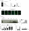
sI/R activates NF-κB in isolated adult mouse cardiomyocytes. A, adult mouse cardiomyocytes underwent 30 min of ischemia followed by reoxygenation for up to 4 h. Details of the sI/R and of intervention are shown. B, sI/R did not induce cell death during the 4-h study period. Cardiomyocytes were exposed to normoxia, ischemia for 30 min, or sI/R for 4 h. Cell death was analyzed by ELISA as described under “Experimental Procedures.” Doxorubicin hydrochloride (Dox.; 1 μm for 24 h) was used as a positive control. *, p < 0.001 versus control (normoxia) and ischemia alone. C, sI/R induced oxidative stress. Oxidative stress was quantified at 4 h after reoxygenation by quantitating the lipid peroxidation products MDA and 4-HNE, as described under “Experimental Procedures.” *, p < 0.001 versus normoxia; n = 6). D, sI/R-mediated oxidative stress was confirmed by DCF fluorescence. Cardiomyocytes loaded with the ROS-sensitive fluorophore dichlorofluorescein diacetate were exposed to ischemia alone or sI/R. Representative fluorescent micrographs at the indicated time periods are shown (n=3). E, sI/R activated NF-κB DNA binding activity. Cardiomyocytes were exposed to sI/R for upto 2 h, and nuclear extracts were assessed for NF-κB DNA binding activity by EMSA, as described under “Experimental Procedures” (n = 3/group). A representative of three independent experiments is shown. Arrow, NF-κB-specific DNA-protein complexes. F, sI/R-mediated NF-κB p65 translocation into the nucleus. Cardiomyocytes underwent sI/R for up to 2 h, and nuclear protein extracts were analyzed for p65 levels by ELISA (n = 3; *, p < 0.05; **, p < 0.01 versus normoxia). G, ROS were generated prior to NF-κB activation. Results from D and E are compared to demonstrate that ROS (oxidative stress) are generated prior to activation of NF-κB in cardiomyocytes following sI/R. *, p < 0.01 versus normoxia; †, p < 0.05; ††, p < 0.001 versus normoxia (n = 3). H, sI/R stimulated NF-κB-driven reporter gene activity. Cardiomyocytes were transduced with adenoviral NF-κB-Luc vector and 24 h later were exposed to sI/R (n = 12). Cotransfection with a β-galactosidase vector (Ad.β-galactosidase) was used to control for transfection efficiency. Ad.MCS-Luc served as a control. Firefly luciferase andβ-galactosidase levels were assayed after 12 h. *, p < 0.001 versus normoxia. I, sI/R induces p65 phosphorylation. Cardiomyocytes were exposed to sI/R for 2 h, and nuclear extracts were analyzed for phospho-p65 levels by immunoblotting. A representative of three independent experiments is shown. Lamin A/C levels demonstrate the purity and equal loading of the nuclear extracts in each lane.
FIGURE 7.
FIGURE 7.
Simulated ischemia/reoxygenation induces NF-κB activation via IKK. A, adenoviral transduction of dnIKKβ blunted sI/R-mediated IKKβ activity. Cardiomyocytes were transfected with Ad.dnIKKβ (100 MOI). Ad.GFP served as a control. After 24 h, cells were exposed to sI/R for 2 h. IKK activity was analyzed by an in vitro kinase assay using glutathione S-transferase-IκBas the substrate (n = 3). Actin served as a loading control. B, sI/R induced IκB-α degradation. Cardiomyocytes were treated as in A but for 1 h and then analyzed for IκB-α degradation by immunoblotting (n = 3). Actin served as a loading control. C, sI/R induced NF-κB activation via IKKβ and IκB-α degradation. Cardiomyocytes were pretreated with the free radical scavenger PDTC (100 μm in PBS for 1 h) or transduced with adenoviral dnIKKβ or dnIκB-α for 24 h, followed by sI/R for 2 h. PBS and Ad.GFP served as controls. NF-κB DNA binding activity was analyzed by EMSA (n = 3). D, the proteasomal inhibitor MG-132 blunted sI/R-mediated NF-κB activation. Cardiomyocytes were pretreated with MG-132 (5 μm in DMSO for 1 h) prior to sI/R for 2 h. DMSO served as a control. NF-κB DNA binding activity was analyzed by EMSA (n = 3). The arrows in C and D indicate NF-κB-specific DNA-protein complexes.
FIGURE 8.
FIGURE 8.
Simulated ischemia/reperfusion induces biologically active IL-18 via IKK-NF-κB signaling. A, PDTC inhibited sI/R-mediated IL-18 mRNA expression. Cardiomyocytes pretreated with PDTC (100 μm in PBS for 1 h) were exposed to sI/R for 4 h, and IL-18 mRNA expression was analyzed by Northern blotting. 28 S rRNA served as a control. A representative of three independent experiments is shown. B, sI/R induced IL-18 mRNA expression via IKK and IκB-α degradation. Cardiomyocytes transduced with adenoviral dnIKKβ, dnIκB-α, or GFP for 24 h were exposed to sI/R for 4 h. IL-18 mRNA expression was analyzed as in A (n = 3). TNF-α (10 ng/ml for 4 h) served as a positive control. C, sI/R-mediated IL-18 expression was independent of TNF-α. Cardiomyocytes were incubated with TNF-α-neutralizing antibodies or control IgG (10 μg/ml for 1 h) prior to sI/R. IL-18 protein levels were analyzed by immunoblotting (n = 3). Actin served as a loading control. D, TNF-α-neutralizing antibodies attenuated TNF-α-induced IL-18 expression. To demonstrate the efficacy of TNF-α-neutralizing antibodies, cardiomyocytes were incubated with αTNF-α antibodies or control IgG (10 μg/ml for 1 h) prior to TNF-α addition (10 ng/ml for 4 h). IL-18 protein levels were quantified by immunoblotting (n = 3). Actin served as a loading control. E, PDTC inhibited sI/R-mediated IL-18 protein expression. Cardiomyocytes treated as in A were analyzed for IL-18 protein expression by immunoblotting (n = 3). Actin served as a control. F, sI/R induced IL-18 protein expression via IKKβ and IκB-α degradation. Cardiomyocytes treated as in B were analyzed for IL-18 protein levels by immunoblotting. TNF-α served as a positive control. G, the proteasomal inhibitor MG-132 and the IKK inhibitor SC-514 inhibited sI/R-mediated IL-18 protein expression. Cardiomyocytes pretreated with MG-132 (5 μm in DMSO for 1 h) or SC-514 (10 μm in DMSO for 1 h) were exposed to sI/R for 4 h. IL-18 protein expression was analyzed by immunoblotting (n = 3). Actin served as a control. H, dnIKK and dnIκB-α blunted TNF-α-induced NF-κB activation. Cardiomyocytes transfected as in B were treated with TNF-α (10 ng/ml for 1 h) and were analyzed for nuclear NF-κB p65 levels by ELISA (n = 3). *, p < 0.001 versus untreated; †, p < 0.01 versus TNF-α. I, dnIKK and dnIκB-α failed to modulate sI/R-mediated JNK phosphorylation. Cardiomyocytes transfected as in B were exposed to sI/R for 2 h. Total and phospho-JNK levels in cleared cell lysates were analyzed by immunoblotting (n = 3). J, sI/R stimulated IL-18 secretion via IKK and IκB-α degradation. Cardiomyocytes were treated as in A and B, and IL-18 levels in culture supernatants were analyzed for IL-18 levels by ELISA (n = 6). TNF-α served as a positive control (10 ng/ml for 4 h). The levels in the treated samples were normalized to the normoxic samples (equal to 1), and the results are expressed as -fold induction from untreated (normoxic). *, p < 0.01 versus untreated; †, p < 0.05 versus sI/R. K, sI/R-stimulated IL-18 was biologically active. Mouse splenocytes (5 × 10⁶/ml) were incubated with and without cardiomyocyte-derived culture supernatants from G and LPS (1 μg/ml) for 24 h at 37 °C. After incubation, supernatants were analyzed for IFN-γ production by ELISA (n = 6). To demonstrate IL-18 specificity, culture supernatants were preincubated with anti-IL-18-neutralizing antibodies (10 μg/ml for 1 h at 37 °C). Normal IgG at similar concentrations served as a control. *, p < 0.001 versus untreated; †, p < 0.05 versus 200 μl of culture supernatant + LPS (n = 6).

References

    1. Okamura, H., Tsutsi, H., Komatsu, T., Yutsudo, M., Hakura, A., Tanimoto, T., Torigoe, K., Okura, T., Nukada, Y., Hattori, K., Akita, K., Namba, M., Tanabe, F., Konishi, K., Fukuda, S., and Kurimoto, M. (1995) Nature 378 88-91 - PubMed
    1. Gracie, J. A., Robertson, S. E., and McInnes, I. B. (2003) J. Leukocyte Biol. 73 213-224 - PubMed
    1. Dinarello, C. A., Novick, D., Puren, A. J., Fantuzzi, G., Shapiro, L., Muhl, H., Yoon, D. Y., Reznikov, L. L., Kim, S. H., and Rubinstein, M. (1998) J. Leukocyte Biol. 63 658-664 - PubMed
    1. Wang, M., Markel, T. A., and Meldrum, D. R. (2008) Shock 30 3-10 - PubMed
    1. Arend, W. P., Palmer, G., and Gabay, C. (2008) Immunol. Rev. 223 20-38 - PubMed

Publication types

MeSH terms