. 2009 Mar 27;284(13):8292-300.
doi: 10.1074/jbc.M805877200. Epub 2009 Jan 21.

N-Glycan Moieties in Neonatal Fc Receptor Determine Steady-state Membrane Distribution and Directional Transport of IgG

Affiliations

N-Glycan Moieties in Neonatal Fc Receptor Determine Steady-state Membrane Distribution and Directional Transport of IgG

Timothy T Kuo et al. J Biol Chem. .

Abstract

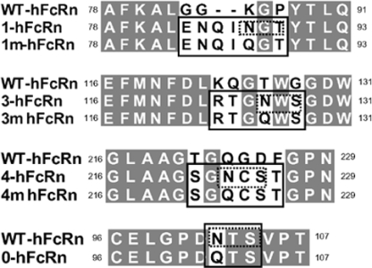
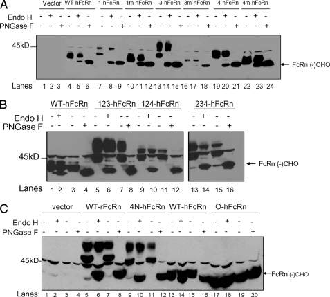
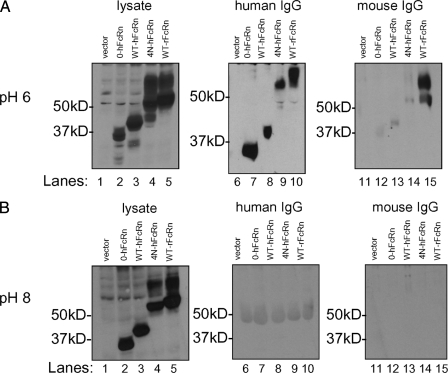
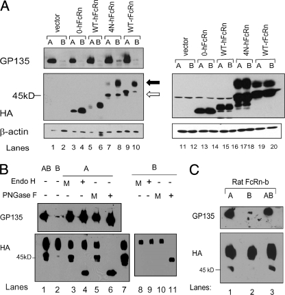
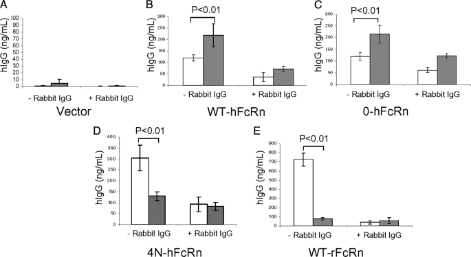
The neonatal Fc receptor (FcRn) is a major histocompatibility complex class I-related molecule known to protect IgG and albumin from catabolism and transport IgG across polarized epithelial cells in a bidirectional manner. Previous studies have shown species-specific differences in ligand binding, IgG transport direction, and steady-state membrane distribution when expressed in polarized epithelial cells. We hypothesized that these differences may be due to the additional N-glycans expressed on the rat FcRn, because N-glycans have been proposed to function as apical targeting signals, and that two of the N-glycan moieties have been shown to contribute to the IgG binding of rat FcRn. A panel of mutant human FcRn variants was generated to resemble the N-glycan expression of rat FcRn in various combinations and subsequently transfected into Madin-Darby canine kidney II cells together with human beta2-microglobulin. Mutant human FcRn clones that contained additional N-glycan side-chain modifications, including that which was fully rodentized, still exhibited specificity for human IgG and failed to bind to mouse IgG. At steady state, the mutant human FcRn with additional N-glycans redistributed to the apical cell surface similar to that of rat FcRn. Furthermore, the rodentized human FcRn exhibited a reversal of IgG transport with predominant transcytosis from an apical-to-basolateral direction, which resembled that of the rat FcRn isoform. These studies show that the N-glycans in FcRn contribute significantly to the steady-state membrane distribution and direction of IgG transport in polarized epithelia.


Figures

FIGURE 1.
FIGURE 1.
Amino acid sequence of hFcRn clones. Amino acid sequence of wild-type (WT) hFcRn and sequence mutations of hFcRn (solid rectangular box), which correspond to that of rFcRn sequence. The predicted glycosylation sequence for rFcRn is highlighted in dashed rectangular box. Corresponding non-glycosylation sequence, wherein conservative substitution of asparagine with glutamine, were created on “mutant” (m) clones: 1m-hFcRn, 3m-hFcRn, 4m-hFcRn, and 0-hFcRn clones.
FIGURE 2.
FIGURE 2.
Functional expression of WT-hFcRn and “rodentized” hFcRn clones. Stable clones of hFcRn and mutant hFcRn clones were generated by transfection of MDCK II cells with hβ2m, and the lysates examined for expression of all transfected clones. Lysate incubation with Endo H removed high mannose N-glycan residues but not complex N-glycan residues, resulting in an upper band (hFcRn with complex N-glycans) and a lower band (hFcRn without any N-glycan residues, deglycosylated FcRn, “FcRn(-)CHO”). PNGase F removed all N-glycan residues, thus resulting in only a single lower band, representing deglycosylated FcRn, “FcRn(-)CHO.” The clones consisted of wild type hFcRn (A, lanes 4–6), hFcRn with one additional N-glycan (A, lanes 7–24), hFcRn with two addition N-glycans (B, lanes 5–16), “Rodentized” hFcRn with three additional N-glycans (C, lanes 9–12), wild-type rat FcRn (C, lanes 13–16), and hFcRn without any N-glycans (C, lanes 17–20).
FIGURE 3.
FIGURE 3.
WT-hFcRn and rodentized hFcRn binds to human IgG but not mouse IgG at pH6. Cells were lysed at CHAPS, pH 6 or pH 8, before incubation with human or mouse IgG and subsequently precipitated with protein G-Sepharose. WT-hFcRn, rodentized hFcRn (4N-hFcRn), and 0-hFcRn were able to bind to human IgG at pH 6 but not pH 8 (A, lanes 7–10; B, lanes 7–10). No binding was seen with mouse IgG at either pH (A, lanes 12–14; B, lanes 12–14). Only WT-rFcRn bound to mouse IgG at pH6 but not pH 8 (A, lane 15; B, lane 15).
FIGURE 4.
FIGURE 4.
Membrane distribution of FcRn. Cells were grown on Transwells and allowed to polarize before surface membrane labeling with biotin at the apical (A), basolateral (B), or both apical and basolateral (AB) cell surface(s) and followed by immunoprecipitation with avidin-agarose. Basolateral predominant membrane distributions of FcRn were observed with 0-hFcRn and WT-hFcRn clones (A, lanes 3–6). Increased apical membrane distribution was observed with 4N-hFcRn and WT-rFcRn (lanes 7–10). In both WT-rFcRn and 4N-hFcRn clones, the high mannose isoform of FcRn was distributed to the apical cell surface (A, lanes 7 and 9, lower bands). Closed arrow indicates mature isoform, and open arrow indicates immature isoform (A). Immunoblotting of total lysate with 12CA5 for FcRn is shown (A, lanes 11–20). Confirmation of membrane bound high mannose and complex N-glycan isoforms in 4N-hFcRn were performed using Endo H, PNGase F, or mock (M) digestion (B). A separate rat FcRn clone (rat FcRn-b) had also been generated (C). Membrane protein isolation from apical (A), basolateral (B), and both apical and basolateral (AB) surfaces demonstrated the presence of rat FcRn at both membrane surfaces. Different from the clone in A (lanes 9 and 10), this clone showed that the mature isoform was visible in both membrane surfaces (C, lanes 1 and 2).
FIGURE 5.
FIGURE 5.
Reversal of human IgG transport with full rodentization of hFcRn. Bidirectional hIgG transport, apical to basolateral (white bars) and basolateral to apical (gray bars), was observed in all clones, except for Vector clone (negative control), which does not express FcRn (A). 0-hFcRn clone exhibited similar basolateral-to-apical direction of hIgG transport (C) as that of WT-hFcRn (B). 4N-hFcRn clone displayed predominate apical-to-basolateral hIgG transport (D), similar to that of WT-rFcRn (E). Addition of rabbit IgG significantly decreased hIgG transport in all clones.

References

    1. Brambell, F. W. (1969) Proc. Nutr. Soc. 28 35–41 - PubMed
    1. Simister, N. E., and Mostov, K. E. (1989) Cold Spring Harbor Symp. Quant. Biol. 54 571–580 - PubMed
    1. Yoshida, M., Masuda, A., Kuo, T. T., Kobayashi, K., Claypool, S. M., Takagawa, T., Kutsumi, H., Azuma, T., Lencer, W. I., and Blumberg, R. S. (2006) Springer Semin. Immunopathol. 28 397–403 - PubMed
    1. Burmeister, W. P., Huber, A. H., and Bjorkman, P. J. (1994) Nature 372 379–383 - PubMed
    1. Rodewald, R., Lewis, D. M., and Kraehenbuhl, J. P. (1983) CIBA Found. Symp. 95 287–299 - PubMed

Publication types

MeSH terms

LinkOut - more resources