. 2009 Apr;23(4):444-53.
doi: 10.1210/me.2008-0106. Epub 2009 Jan 22.

Susceptibility to autoimmunity and B cell resistance to apoptosis in mice lacking androgen receptor in B cells

Affiliations

Susceptibility to autoimmunity and B cell resistance to apoptosis in mice lacking androgen receptor in B cells

Saleh Altuwaijri et al. Mol Endocrinol. 2009 Apr.

Erratum in

Abstract

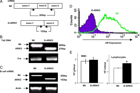
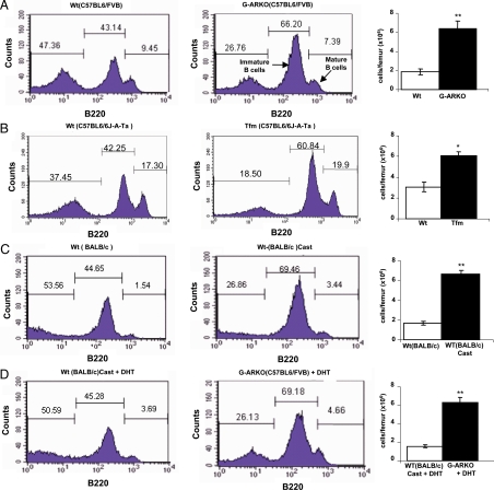
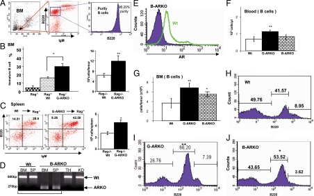
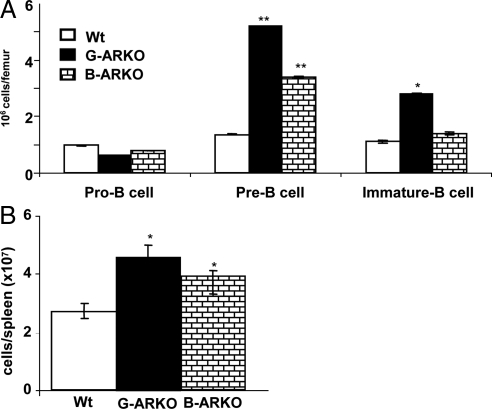
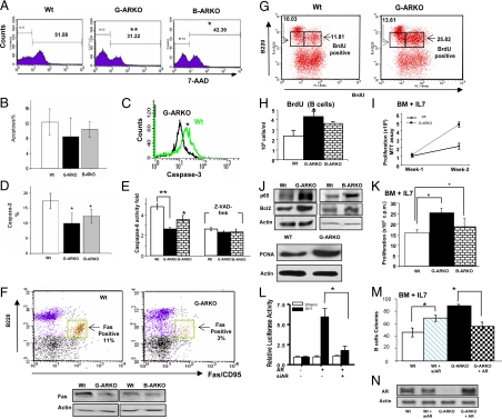
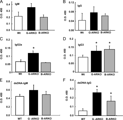
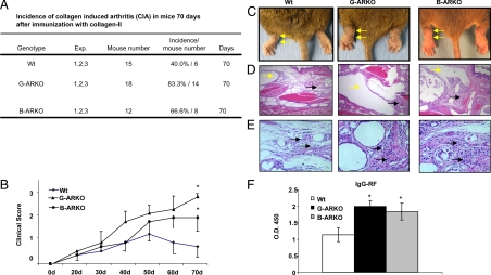
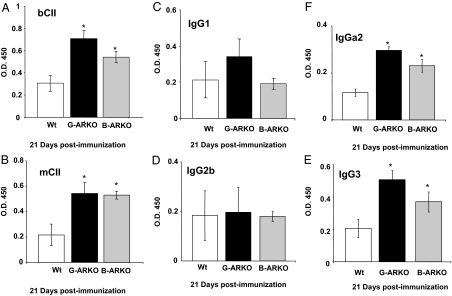
Estrogens have been linked to a higher female incidence of autoimmune diseases. The role of androgen and the androgen receptor (AR) in autoimmune diseases, however, remains unclear. Here we report that the lack of AR in B cells in different strains of mice, namely general AR knockout, B cell-specific AR knockout, and naturally occurring testicular feminization mutation AR-mutant mice, as well as castrated wild-type mice, results in increased B cells in blood and bone marrow. Analysis of the targeted mice, together with bone marrow transplantation using Rag1(-/-) recipients, overexpression of retrovirally encoded AR-cDNA, and small interfering RNA-mediated AR mRNA knockdown approaches also show that the B cell expansion results from resistance to apoptosis and increased proliferation of bone marrow precursor B cells, accompanied by changes in several key modulators related to apoptosis, such as Fas/FasL signals, caspases-3/-8, nuclear factor-kappaB, and Bcl-2. We also show that the effects of AR loss are, in part, B cell intrinsic. Mice bearing AR-deficient B cells show increased levels of serum IgG2a and IgG3 as well as basal double-stranded DNA-IgG antibodies and are more vulnerable to development of collagen-induced arthritis. Together, these data indicate that androgen/AR play a crucial role in B cell homeostasis and tolerance. Therapies targeting AR might provide an alternative strategy with which to battle autoimmune diseases.


Figures

Figure 1
Figure 1
Characterization of G-ARKO mice (A) G-ARKO (C57BL6/FVB) mice, bearing a deletion of the AR DNA binding domain (exon 2). B, Genotyping of tissue DNA isolated from G-ARKO and Wt littermates using “select and 2–9” primers; 600 bp represents Wt AR and 270 bp represents G-ARKO. C, We used the primers 5′-AATGGGACCTTGGATGGAGAAC-3′ and 5′-TCCCTGCTTCATAACATTTCCG-3′ for RT-PCR, to obtain an AR transcript of 305 bp from Wt AR, but only 153 bp when exon 2 is deleted. Full-length AR mRNA is not expressed in B cells from the bone marrow of G-ARKO mice. D, FACS analysis shows intracellular AR protein is not detected in B cells from the bone marrow of G-ARKO mice. E, Automated blood analysis shows peripheral blood lymphocytes, but not total white blood cells (WBC), are increased in G-ARKO (n = 24) compared with Wt (n =18). Data are mean ± sd. *, P < 0.05.
Figure 2
Figure 2
Expansion of developing B cells in distinct strains of mice lacking AR or with ablation of AR function after castration. A, High numbers of precursor B220low B cells in G-ARKO (C57BL6/FVB) (n = 5) compared with Wt (C57BL6/FVB) mice (n = 5). The absolute numbers of the B cells from BM (106 cells per femur) are increased in the G-ARKO mice (far right). B, High numbers of precursor B220low B cells in Tfm (C57B6/6J-A-Ta) (n = 4) compared with Wt mice (C57B6/6J-A-Ta) (n = 4). C, High numbers of precursor B220low B cells in castrated BALB/c (n = 5) compared with Wt BALB/c mice (n = 5). D, DHT supplementation reduces B cells in bone marrow of castrated BALB/c mice (n = 5) but not in G-ARKO mice (n = 5), suggesting that AR mediates this effect. Cast, Castration.
Figure 3
Figure 3
Absence of AR in B cells results in an expansion of B cells in bone marrow (BM) and spleen. A, Purification and sorting the immature B cell (B220+ IgM) cells for B cell BM transplantation. B, Distribution and absolute number of BM immature B cells in Rag1−/− recipients transplanted with BM cells from Wt (n = 5) and G-ARKO mice (n = 5). C, FACS analysis and absolute numbers of B cells in spleens of RAG1−/− recipient transplanted with BM cells from Wt (n = 5) and G-ARKO mice (n = 5). D, Genotyping of different tissue B cell DNA isolated from B-ARKO and Wt mice using “select and 2–9” primers, specific for sequences flanking the AR gene. Wt AR yields a PCR product of 600 bp, and the conditionally deleted region in the B-ARKO gene yields a fragment of 270 bp. B cells from B-ARKO BM and spleen (SP) only display the short AR 270 bp, suggesting specific ablation of the AR in B cells. E, Intracellular staining of AR in precursor B220low B cells (gated by FACS analysis) from BM of Wt and B-ARKO mice. Blood (panel F) and BM analyses (panel G) show expansion of B cells in G-ARKO (n = 18) mice as compared with those of Wt mice (n = 18). B-ARKO (n = 7) mice show expansion of B cells in BM but not in the blood. H–J, Distribution of B lymphocytes in the BM of Wt, G-ARKO, and B-ARKO mice. Data are mean ± sd. *, P < 0.05; **, P < 0.01. TH, Thymus; KD, kidney.
Figure 4
Figure 4
Stage of differentiation of B220+ cells in bone marrow and in periphery of G-ARKO, B-ARKO. A, Bone marrow cells from G-ARKO, B-ARKO, and Wt mice were analyzed for cell surface marker expression patterns using flow cytometry with markers B220, CD2, and IgM. Three populations of B220+ gated cells were observed: pro-B cells (B220+, CD2, IgM), pre-B cells (B220+, CD2+, IgM) and immature B cells (B220+,CD2+, IgMlow). B, The subset distribution of B cells (B220+), in the spleens of G-ARKO, B-ARKO, and Wt mice.
Figure 5
Figure 5
G-ARKO B cells are resistant to apoptosis. A, Bone marrow (BM) immature B cells from G-ARKO and B-ARKO mice were more resistant to apoptosis than cells from Wt littermates. Total BM cells were cultured in vitro for 14 d in MesenCult medium supplemented with IL-7 and then stained with fluorochrome-conjugated anti-B220, anti-IgM, and 7-AAD and analyzed by flow cytometry. Histograms show 7-AAD staining profiles of gated B220low IgMlow immature B cells. B, Total BM cells were cultured in vitro for 48 h in MesenCult medium supplemented with IL-7. The TUNEL assay was used to detect apoptotic cells, with these events gated on B220low. C and D, Caspase-3 activity was analyzed in B cells from Wt and G-ARKO BM cultures cultured in vitro for 14 d in MesenCult medium supplemented with IL-7, stained with fluorochrome-conjugated anti-B220 and anti-caspase-3, and then analyzed by FACS gating on B220low developing B cells. E, Caspase-8 analysis of immature B cells from Wt, G-ARKO, and B-ARKO BM cells cultured in vitro for 14 d in MesenCult medium containing IL-7. We analyzed levels of caspase-8 in BM B cell lysates by ELISA and used general caspase inhibitor Z-VAD-fmak to reduce caspase-8 activity. F, Distribution of Fas (CD95)-positive B220+lowcells in Wt and G-ARKO BM cells cultured in vitro for 14 d in MesenCult medium containing IL-7 and then stained with fluorochrome-conjugated anti-B220 and anti-CD95 (Fas). Flow cytometry analysis and Western blot revealed that BM B220low cells from G-ARKO, B-ARKO mice have less Fas-positive cells than Wt littermates. G, BrdU staining of B cells from G-ARKO and Wt littermates. G-ARKO and Wt littermates were injected ip with 1 mg of BrdU twice a day for 4 d. BM cells were stained with fluorochrome-conjugated anti-B220 and anti-BrdU and analyzed by FACS for B220low BrdU+ cells. H, The absolute number of BrdU-positive B220low cells was increased in G-ARKO (n = 9) and B-ARKO (n = 6) compared with Wt mice (n = 9). I, Proliferation analysis for bone marrow B cells from Wt and G-ARKO littermates in the presence of 20 nm IL-7. J, Western blot analysis of NF-kB/p65 and Bcl2 from BM B cell lysates of Wt, G-ARKO, and B-ARKO mice littermates. K, BM B cell proliferation was measured by [3H]thymidine incorporation in the presence of 20 nm IL-7 (see Materials and Methods). Thymidine incorporation is greater in immature B cells from G-ARKO (n = 5) and B-ARKO mice (n = 5) compared with Wt (n = 5). L, AR activity is knocked down by AR siRNA and restored by AR-cDNA. CV-1 cells were cotransfected with ARE4-Luc, pBabe-AR (or pBabe vector), and AR-siRNA using SuperFect. phRL-TK was cotransfected as the control for normalization. Total plasmid DNA amounts were balanced by empty vectors. After 24 h transfection, cells were treated with or without 10 nm DHT. After 16–18 h incubation, cells were harvested for the luciferase reporter assay. Each Luc activity is presented relative to the transactivation observed in the absence of DHT and is the mean ± sd of four experiments. M, We plated BM cells infected with retroviruses harboring pBabe-AR, pSuperior-ARsiRNA, and vectors in methylcellulose-containing media supplemented with IL-7. Colonies containing more than 30 cells were counted after 14 d. AR knockdown in Wt BM cells by siRNA induces colony forming, and AR restoration in G-ARKO BM cells reduces colony forming. N, Western blot revealed AR restored by AR-cDNA in G-ARKO BM cells. Data are presented as mean ± sd *, P < 0.05; **, P < 0.01. PCNA, Proliferating cell nuclear antigen.
Figure 6
Figure 6
Levels of Ig isotypes by ELISAs. Relative serum concentrations of (A) IgM, (B) IgG, (C) IgG2a, (D) IgG3, (E) dsDNA-IgM, and (F) dsDNA-IgG were higher in G-ARKO mice compared with Wt mice. IgG, IgG3, and dsDNA-IgM are higher in B-ARKO mice than in Wt mice. The data were obtained from six pairs of mice. Data are presented as mean ± sd. *, P < 0.05.
Figure 7
Figure 7
G-ARKO and B-ARKO mice are more vulnerable to CIA than their Wt littermates. A, Incidence of CIA in mice 70 d after immunization with CII. B, Average clinical scores per paw during disease progression. C, External joint phenotype after inducing arthritis is indicated by arrows. D, Histological features of joint sections. Yellow arrows indicate extended synovial bursa. Black arrows indicate hypertrophic synovial membrane. E, Infiltration of lymphocytes in synovial membrane of joints after CIA (arrows). F, Relative serum IgG-RF levels at the end of the experiment were increased in G-ARKO (n = 5) and B-ARKO (n = 3), but not in Wt mice (n = 5). Data, mean ± sd; *, P < 0.05; **, P < 0.01.
Figure 8
Figure 8
Measurement of CII-specific Ab by ELISA after immunization. Sera from G-ARKO, B-ARKO, and Wt mice. Mice were immunized with collagen II, and after 21 d the sera were collected and analyzed for the presence of total anti-body of (A) bCII (bCII-specific), (B) mCII (mCII-specific), and (C–F) analysis of isotype expression in the mCII-specific Ab response. mCII-specific isotypes were quantitated using the sera described in panel A and a solid phase ELISA in which the detection Abs were specific for the isotypes listed. The quantity of Ab detected is expressed as relative OD450 based on a CII-specific reference serum. Data are presented as mean ± sd. *, P < 0.05.

References

    1. Chang CS, Kokontis J, Liao ST 1988 Molecular cloning of human and rat complementary DNA encoding androgen receptors. Science 240:324–326 - PubMed
    1. Heinlein CA, Chang C 2004 Androgen receptor in prostate cancer. Endocr Rev 25:276–308 - PubMed
    1. Tsai MJ, O'Malley BW 1994 Molecular mechanisms of action of steroid/thyroid receptor superfamily members. Annu Rev Biochem 63:451–486 - PubMed
    1. Rochette-Egly C 2005 Dynamic combinatorial networks in nuclear receptor-mediated transcription. J Biol Chem 280:32565–32568 - PubMed
    1. Abu EO, Horner A, Kusec V, Triffitt JT, Compston JE 1997 The localization of androgen receptors in human bone. J Clin Endocrinol Metab 82:3493–3497 - PubMed

Publication types

MeSH terms