. 2009 Apr;329(1):335-41.
doi: 10.1124/jpet.108.143453. Epub 2009 Jan 22.

Substrate selectivity of 5-hydroxyeicosanoid dehydrogenase and its inhibition by 5-hydroxy-Delta6-long-chain fatty acids

Affiliations

Substrate selectivity of 5-hydroxyeicosanoid dehydrogenase and its inhibition by 5-hydroxy-Delta6-long-chain fatty acids

Pranav Patel et al. J Pharmacol Exp Ther. 2009 Apr.

Abstract

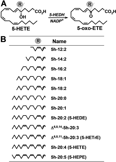
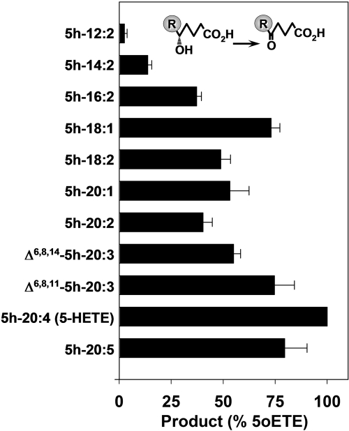
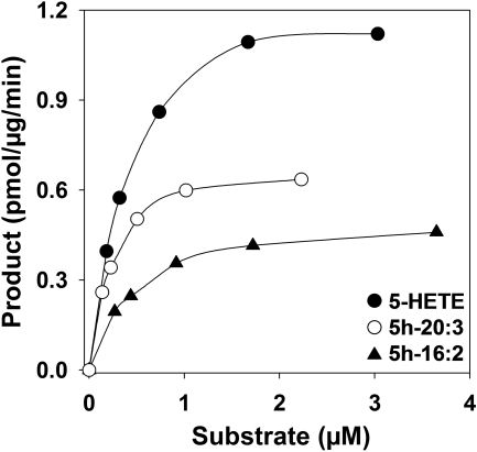
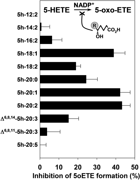
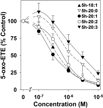
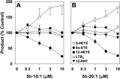
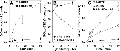
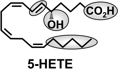
5-Oxo-6E,8Z,11Z,14Z-eicosatetraenoic acid (5-oxo-ETE) is a metabolite of the 5-lipoxygenase (5-LO) product 5S-hydroxy-6E,8Z,11Z,14Z-eicosatetraenoic acid (5-HETE), formed by the microsomal enzyme 5-hydroxyeicosanoid dehydrogenase (5-HEDH). 5-oxo-ETE is a chemoattractant for neutrophils and eosinophils, both in vitro and in vivo. To examine the substrate selectivity of 5-HEDH and to search for potential inhibitors, we prepared a series of 5S-hydroxy fatty acids (C(12) to C(20) containing zero to four double bonds) by total chemical synthesis and examined their metabolism by microsomes from monocytic U937 cells. Although most of these fatty acids were oxidized to their 5-oxo metabolites by 5-HEDH, 5-HETE seemed to be the best substrate. However, substrates containing less than 16 carbons, a methylated alpha-carboxyl group, or a hydroxyl group at the omega-end of the molecule were not substantially metabolized. Some of the fatty acids tested were fairly potent inhibitors of the formation of 5-oxo-ETE by 5-HEDH, in particular 5-hydroxy-6-octadecenoic acid and 5-hydroxy-6-eicosenoic acid. Both substances selectively inhibited 5-oxo-ETE formation by human peripheral blood mononuclear cells incubated with arachidonic acid and calcium ionophore without affecting the formation of leukotriene B(4), 12-HETE, or 12-hydroxy-5,8,10-heptadecatrienoic acid. We conclude that the requirements for appreciable metabolism by 5-HEDH include a chain length of at least 16 carbons, a free alpha-carboxyl group, and a hydrophobic group at the omega-end of the molecule. 5-Hydroxy-Delta(6) C(18) and C(20) fatty acids selectively inhibit 5-HEDH without inhibiting 5-LO, leukotriene A(4) hydrolase, 12-lipoxygenase, or cyclooxygenase. Such compounds may be useful in defining the role of 5-oxo-ETE and its mechanism of synthesis.


Figures

Fig. 1.
Fig. 1.
Metabolism of 5-hydroxy fatty acids by 5-HEDH. A, conversion of 5-HETE to 5-oxo-ETE by 5-HEDH. B, structures of 5S-hydroxy fatty acids that were tested for substrate and/or inhibitor activity with respect to microsomal 5-HEDH.
Fig. 2.
Fig. 2.
Metabolism of 5-hydroxy fatty acids by microsomal 5-HEDH. Microsomal fractions (15 μg/ml) from PMA-differentiated U937 cells were incubated with 5-hydroxy fatty acids (3 μM) in the presence of NADP+ (100 μM) for 10 min at 37°C. The amounts of 5-oxo products formed were determined by RP-HPLC (Table 1). The values are means ± S.E. of four independent experiments.
Fig. 3.
Fig. 3.
Effects of substrate concentration on the amounts of 5-oxo products formed by microsomal 5-HEDH. Microsomal fractions from PMA-differentiated U937 cells were incubated with different concentrations of 5h-16:2 (▴), Δ6,8,11-5h-20:3 (○), or 5-HETE (•) as described in the legend to Fig. 2, and the amounts of 5-oxo products formed were determined by RP-HPLC (Table 1). The values are from a single experiment and are representative of five independent experiments with similar results.
Fig. 4.
Fig. 4.
Inhibition of 5-HEDH by 5-hydroxy fatty acids. Microsomal fractions (15 μg/ml) from PMA-differentiated U937 cells were incubated with 5-HETE (1 μM) in the presence of various 5-hydroxy fatty acids (0.3 μM) and NADP+ (100 μM) for 10 min at 37°C. The amounts of 5-oxo-ETE formed were determined by RP-HPLC. The values are means ± S.E. of four independent experiments.
Fig. 5.
Fig. 5.
Concentration-response curves for the inhibition by 5-hydroxy fatty acids of 5-oxo-ETE formation by microsomal 5-HEDH. Microsomal fractions (15 μg/ml) from PMA-differentiated U937 cells were preincubated with various concentrations of 5h-18:1 (▴), 5h-20:1 (•), 5h-20:2 (○), 5h-20:0 (▿), and Δ6,8,11-5h-20:3 (5h-20:3, ▾) for 5 min followed by incubation with 5-HETE (1 μM) in the presence of NADP+ (100 μM) for 10 min. The amounts of 5-oxo-ETE formed were determined by RP-HPLC. The values are means ± S.E. (n = 4).
Fig. 6.
Fig. 6.
Effects of 5h-18:1 and 5h-20:1 on the formation of AA metabolites by human PBMCs. PBMCs (5 × 10⁶ cells in 1 ml) were preincubated for 1 min with either 5h-18:1 (n = 6) (A) or 5h-20:1 (n = 4) (B) and then incubated with AA (20 μM), A23187 (5 μM), and tBuOOH (100 μM) for 10 min. The amounts of 5-HETE (○), 12-HETE (▪), 12-HHT (▾), LTB4 (▵), and 5-oxo-ETE (•) were analyzed by precolumn extraction/RP-HPLC as described under Materials and Methods using a linear gradient prepared from solvents A (water), B (acetonitrile), and C (methanol), all containing 0.02% acetic acid, as follows: 0 min, 46.5% A, 28.5% B, 25% C; and 24 min, 11.1% A, 37.6% B, 51.3% C at a flow rate of 1 ml/min. All values are means ± S.E.
Fig. 7.
Fig. 7.
Importance of the α and ω ends of the substrate for metabolism by 5-HEDH. A, time courses for the metabolism of 5-HETE (2 μM) to 5-oxo-ETE (□) and 5-HETE-Me (2 μM) to 5-oxo-ETE-Me (•) by microsomal fractions (30 μg/ml protein) from human neutrophils in the presence of NADP+ (100 μM). B, concentration-response relationships for the effects of 5-HETE-Me (•) and 5,18-diOH-18:2 (▵) on the conversion of 5-HETE (1 μM) to 5-oxo-ETE by neutrophil and U937 cell microsomes, respectively, in the presence of NADP+ (100 μM). Microsomal fractions (30 μg/ml protein) were incubated with 5-hydroxy fatty acids for 15 min. C, time courses for the metabolism of 5-HETE to 5-oxo-ETE (□), 5h-18:2 to 5-oxo-18:2 (○), and 5,18-diOH-18:2 to 5-oxo-18-HODE (•) by microsomal fractions (30 μg/ml protein) from PMA-differentiated U937 cells in the presence of NADP+ (100 μM). All values are means ± S.E. (n = 4).
Fig. 8.
Fig. 8.
Structure of 5-HETE, showing the regions of the molecule that are important for metabolism by 5-HEDH.

References

    1. Borgeat P and Samuelsson B (1979) Arachidonic acid metabolism in polymorphonuclear leukocytes: unstable intermediate in formation of dihydroxy acids. Proc Natl Acad Sci U S A 76 3213-3217. - PMC - PubMed
    1. Cossette C, Patel P, Anumolu JR, Sivendran S, Lee GJ, Gravel S, Graham FD, Lesimple A, Mamer OA, Rokach J, et al. (2008) Human neutrophils convert the sebum-derived polyunsaturated fatty acid sebaleic acid to a potent granulocyte chemoattractant. J Biol Chem 283 11234-11243. - PMC - PubMed
    1. Dallaire MJ, Ferland C, Pagé N, Lavigne S, Davoine F, and Laviolette M (2003) Endothelial cells modulate eosinophil surface markers and mediator release. Eur Respir J 21 918-924. - PubMed
    1. Erlemann KR, Cossette C, Grant GE, Lee GJ, Patel P, Rokach J, and Powell WS (2007) Regulation of 5-hydroxyeicosanoid dehydrogenase activity in monocytic cells. Biochem J 403 157-165. - PMC - PubMed
    1. Erlemann KR, Rokach J, and Powell WS (2004) Oxidative stress stimulates the synthesis of the eosinophil chemoattractant 5-oxo-6,8,11,14-eicosatetraenoic acid by inflammatory cells. J Biol Chem 279 40376-40384. - PubMed

Publication types

MeSH terms

Substances

LinkOut - more resources