

A peroxide bridge between Fe and Cu ions in the O2 reduction site of fully oxidized cytochrome c oxidase could suppress the proton pump
- PMID: 19164527
- PMCID: PMC2650126
- DOI: 10.1073/pnas.0806391106
A peroxide bridge between Fe and Cu ions in the O2 reduction site of fully oxidized cytochrome c oxidase could suppress the proton pump
Abstract
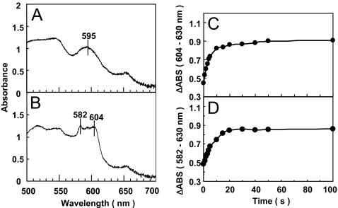
The fully oxidized form of cytochrome c oxidase, immediately after complete oxidation of the fully reduced form, pumps protons upon each of the initial 2 single-electron reduction steps, whereas protons are not pumped during single-electron reduction of the fully oxidized "as-isolated" form (the fully oxidized form without any reduction/oxidation treatment) [Bloch D, et al. (2004) The catalytic cycle of cytochrome c oxidase is not the sum of its two halves. Proc Natl Acad Sci USA 101:529-533]. For identification of structural differences causing the remarkable functional difference between these 2 distinct fully oxidized forms, the X-ray structure of the fully oxidized as-isolated bovine heart cytochrome c oxidase was determined at 1.95-A resolution by limiting the X-ray dose for each shot and by using many (approximately 400) single crystals. This minimizes the effects of hydrated electrons induced by the X-ray irradiation. The X-ray structure showed a peroxide group bridging the 2 metal sites in the O(2) reduction site (Fe(3+)-O(-)-O(-)-Cu(2+)), in contrast to a ferric hydroxide (Fe(3+)-OH(-)) in the fully oxidized form immediately after complete oxidation from the fully reduced form, as has been revealed by resonance Raman analyses. The peroxide-bridged structure is consistent with the reductive titration results showing that 6 electron equivalents are required for complete reduction of the fully oxidized as-isolated form. The structural difference between the 2 fully oxidized forms suggests that the bound peroxide in the O(2) reduction site suppresses the proton pumping function.
Conflict of interest statement
The authors declare no conflict of interest.
Figures
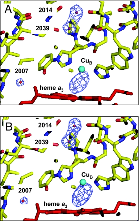
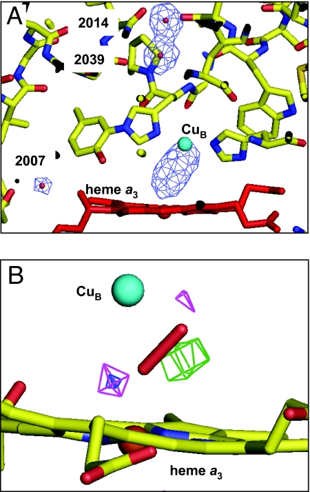
 O distance. Cages are depicted at the 3.8σ level. Heme a3 is depicted by a ball-and-stick model. Carbon, nitrogen, and oxygen atoms are yellow, blue, and red, respectively. The peroxide anion is illustrated by a stick model. Fea3 and CuB are represented in brown and green. The map of the 1.6-Å constraint (pink) has residual density peaks at both ends of the OO bond, whereas the map of the 1.8-Å constraint (green) has a large residual density at the middle of the OO bond. The cages for the 1.7-Å constraint (blue) have the smallest residual density.
References
-
- Ferguson-Miller S, Babcock GT. Heme/copper terminal oxidases. Chem Rev. 1996;96:2889–2908. - PubMed
-
- Moody AJ, Cooper CE, Rich PR. Characterisation of ‘fast’ and ‘slow’ forms of bovine heart cytochrome-c oxidase. Biochim Biophys Acta. 1991;1059:189–207. - PubMed
-
- Thörnström PE, Nilsson T, Malmström BG. The possible role of the closed–open transition in proton pumping by cytochrome c oxidase: The pH dependence of cyanide inhibition. Biochim Biophys Acta. 1988;935:103–108. - PubMed
-
- Mochizuki M, et al. Quantitative reevaluation of the redox active sites of crystalline bovine heart cytochrome c oxidase. J Biol Chem. 1999;274:33403–33411. - PubMed
Publication types
MeSH terms
Substances
Associated data
- Actions
LinkOut - more resources
Full Text Sources
Medical
Molecular Biology Databases
