. 2009 Feb 3;106(5):1512-7.
doi: 10.1073/pnas.0809559106. Epub 2009 Jan 21.

Characterization of the thymic IL-7 niche in vivo

Affiliations

Characterization of the thymic IL-7 niche in vivo

Nuno L Alves et al. Proc Natl Acad Sci U S A. .

Abstract

The thymus represents the "cradle" for T cell development, with thymic stroma providing multiple soluble and membrane cues to developing thymocytes. Although IL-7 is recognized as an essential factor for thymopoiesis, the "environmental niche" of thymic IL-7 activity remains poorly characterized in vivo. Using bacterial artificial chromosome transgenic mice in which YFP is under control of IL-7 promoter, we identify a subset of thymic epithelial cells (TECs) that co-express YFP and high levels of Il7 transcripts (IL-7(hi) cells). IL-7(hi) TECs arise during early fetal development, persist throughout life, and co-express homeostatic chemokines (Ccl19, Ccl25, Cxcl12) and cytokines (Il15) that are critical for normal thymopoiesis. In the adult thymus, IL-7(hi) cells localize to the cortico-medullary junction and display traits of both cortical and medullary TECs. Interestingly, the frequency of IL-7(hi) cells decreases with age, suggesting a mechanism for the age-related thymic involution that is associated with declining IL-7 levels. Our temporal-spatial analysis of IL-7-producing cells in the thymus in vivo suggests that thymic IL-7 levels are dynamically regulated under distinct physiological conditions. This IL-7 reporter mouse provides a valuable tool to further dissect the mechanisms that govern thymic IL-7 expression in vivo.


Conflict of interest statement

The authors declare no conflict of interest.

Figures
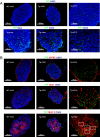
Fig. 1.
Fig. 1.
Thymic IL-7hi cells are of non-hematopoietic origin and lack classical fibroblast and endothelial markers. Phenotypic characterization of IL-7hi cells in fetal thymus. (A) Immunohistochemical analysis of BAC.IL-7.YFP transgenic (Tg) and control (WT) fetal thymus at E14.5 and E17.5. (B) Phenotypic characterization at E14.5 for WT and Tg. Sections were double or triple stained with the indicated Abs. YFP, green; DAPI and CD45, blue; ERTR7, CD31, and MHC class II, red. White squares indicate IL-7hi cells that co-express MHC class II. Data are representative of two or three experiments.
Fig. 2.
Fig. 2.
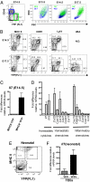
IL-7hi cells display traits of TECs and transcribe homeostatic cytokines and chemokines important for thymocyte development. Flow cytometric analysis of fetal (E13.5, E14.5, and E17.5) and neonatal thymic IL-7hi cells from BAC.IL-7.YFP transgenic mice (Tg) following stromal isolation. (A) Thymic IL-7hi cells comprise a subset of stromal (CD45) cells. The size of IL-7hi cells (green) was compared with that of CD45+ cells (blue). (B) IL-7hi cells were characterized phenotypically for the indicated TEC surface markers. Analysis was performed on thymic stromal cells (CD45 gate). (A,B) Numbers indicate the percentage of each correspondent boxed gate. (N.D., not detectable.) Data are representative of two or three experiments. (C,D) Transcriptional analysis was performed in sorted fetal thymic MHCII+YFP+ and MHCIIYFP stromal (CD45) populations from BAC.IL-7.YFP fetuses (E14.5). (C) Il7 transcripts are markedly up-regulated in fetal thymic IL-7hi cells. The relative expression of Il7 mRNA levels was assessed by quantitative PCR and normalized to Hprt. Thymic MHCII+YFP+ cells were compared relative to MHCIIYFP cells, in which Il7 levels were set to 1. Depicted is the average difference (in fold) between the two cellular subsets from six independent experiments. (D) The relative mRNA expression for 34 cytokines and 37 chemokines was assessed in fetal thymic IL-7hi cells by RT-PCR array. The mRNA level for each target gene was normalized to three house-keeping genes (Hspcb, Gapdh, and Actb). Depicted is the average difference (in fold) between MHCII+YFP+ and MHCIIYFP cells from two independent experiments. (E,F) Il7 transcripts are markedly up-regulated in newborn thymic IL-7hi TECs. (E) Analysis was performed on thymic stroma cells (CD45 gate). (F) The relative expression of Il7 mRNA levels was assessed by qPCR (Right) and normalized to Hprt in sorted thymic MHCII+YFP and MHCII+YFP+ cells and compared relative to MHCIIYFP cells, in which Il7 levels were set to 1. Depicted is the average difference (in fold) between the two cellular subsets from three independent experiments.
Fig. 3.
Fig. 3.
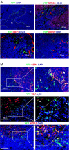
Adult thymic IL-7hi cells are positioned at cortico-medullary regions and bear markers of cortical and medullary TECs. Immunohistochemical analysis of adult thymus (6–8 weeks) of BAC.IL-7.YFP transgenic mice. Sections were double or triple stained with the indicated Ab combination (C, cortex; M, medulla). (A) IL-7hi cells are of nonhematopoietic origin, and predominantly positioned at the cortico-medullary junction and do not costain with endothelial or fibroblast markers [YFP, green; DAPI and CD45 blue; MTS33 (ThB), ERTR7, CD31, red]. (B) IL-7hi cells display markers of cTECs or mTECs. Arrows indicate IL-7hi cells that either coexpress cTEC (CDR1 and Ly51) or mTEC (MTS10) markers (eYFP, green; DAPI, Ly51, blue; CDR1, UEA, MTS10, red). Data are representative of 2–3 experiments.
Fig. 4.
Fig. 4.
The frequency of thymic IL-7hi TECs declines with age. Flow cytometric analysis of IL-7hi TECs isolated from BAC.IL-7.YFP transgenic thymus at different ages. (A) Number of CD45+ cells. (B and C) Number and percentage of stromal (CD45) MHCII+YFP+ cells (gray bars) and MHCII+YFP cells (black bars). (For all time points, n = 7–9, except +6 months, n = 3.) Differences were significant when 2-week and 3- to 6-month groups were compared (CD45+ and MHCII+YFP+, P = 0.0003; MHCII+YFP, P = 0.0037). (D) Flow cytometric analysis showing a representative young, adult, and old thymus (gating on CD45 cells). Numbers indicate the percentage of the correspondent boxed gates.

Similar articles

Cited by

References

    1. Jameson SC, Hogquist KA, Bevan MJ. Positive selection of thymocytes. Annu Rev Immunol. 1995;13:93–126. - PubMed
    1. Kyewski B, Klein L. A central role for central tolerance. Annu Rev Immunol. 2006;24:571–606. - PubMed
    1. Anderson G, Moore NC, Owen JJ, Jenkinson EJ. Cellular interactions in thymocyte development. Annu Rev Immunol. 1996;14:73–99. - PubMed
    1. Derbinski J, Schulte A, Kyewski B, Klein L. Promiscuous gene expression in medullary thymic epithelial cells mirrors the peripheral self. Nat Immunol. 2001;2:1032–1039. - PubMed
    1. Petrie HT, Zuniga-Pflucker JC. Zoned out: functional mapping of stromal signaling microenvironments in the thymus. Annu Rev Immunol. 2007;25:649–679. - PubMed

Publication types

LinkOut - more resources