. 2009 Jan 27;106(4):1169-74.
doi: 10.1073/pnas.0811274106. Epub 2009 Jan 21.

Induction of TLR4-target genes entails calcium/calmodulin-dependent regulation of chromatin remodeling

Affiliations

Induction of TLR4-target genes entails calcium/calmodulin-dependent regulation of chromatin remodeling

Dazhi Lai et al. Proc Natl Acad Sci U S A. .

Abstract

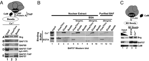
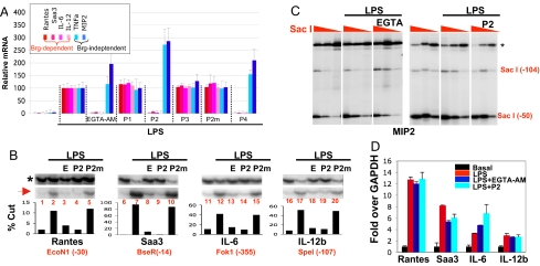
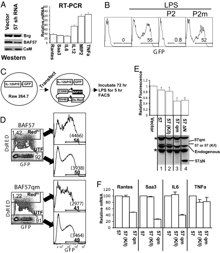
Upon toll-like receptor 4 (TLR4) signaling in macrophages, the mammalian Swi/Snf-like BAF chromatin remodeling complex is recruited to many TLR4 target genes where it remodels their chromatin to promote transcription. Here, we show that, surprisingly, recruitment is not sufficient for chromatin remodeling; a second event, dependent on calcium/calmodulin (CaM), is additionally required. Calcium/CaM directly binds the HMG domain of the BAF57 subunit within the BAF complex. Calcium/CaM antagonists, including a CaM-binding peptide derived from BAF57, abolish BAF-dependent remodeling and gene expression without compromising BAF recruitment. BAF57 RNAi and BAF57 dominant negative mutants defective in CaM binding similarly impair the induction of BAF target genes. Our data implicate calcium/CaM in TLR4 signaling, and reveal a previously undescribed, recruitment-independent mode of regulation of the BAF complex that is probably achieved through a direct CaM-BAF interaction.


Conflict of interest statement

The authors declare no conflict of interest.

Figures

Fig. 1.
Fig. 1.
CaM binds the BAF complex. (A) CaM beads pulled down endogenous BAF complex in addition to affinity-tagged BAF complex from crude HeLa NEs. The integrity of the tag, fused to BAF57, was confirmed by Western blotting using an HRP-conjugated IgG that recognizes the protein A moiety of the tag (blot D). (B) CaM-BAF interactions are calcium-dependent and direct. CaM beads were mixed with 50 μg of HeLa NEs (lanes 2–10) or 0.5 μg of purified BAF complex (lanes 11–13) in the presence of CaCl2 (Upper), EGTA (Lower), or high concentrations of BSA that distorted the gels (lanes 5–13); the BAF complex was affinity-purified by using M2 beads as described (20). CaM beads were washed under the binding condition and eluted with EGTA, and the samples probed with anti-BAF57. (C) M2-beads, which recognize Flag-tagged BAF complex, pulled down endogenous CaM. 1% and 0.3% of NE in the immunoprecipitation (IP) reaction (lanes 1–2) and 50% of precipitated material (lanes 3–4) were loaded on the gel. The IP reaction contained either 5 mM EGTA (lane 3) or 2 mM CaCl2 (lane 4), the latter appearing to impair the IP efficiency.
Fig. 2.
Fig. 2.
CaM binds the HMG domain of BAF57. (A) In vitro cross-linking assay. CaM was cross-linked to the BAF complex and the reaction products resolved by SDS/PAGE before being probed sequentially with antibodies against CaM (Left) and BAF57 (Right). Brackets denote crosslinking between CaM and unknown proteins (Left), or between BAF57 and its neighboring subunits (Right). Arrow, crosslinking between CaM and BAF57. Asterisks, nonspecific immunoreactivity. (B) Anti-BAF57 Western blotting showing that CaM beads captured BAF57 (WT) and the BAF57 point mutant defective in DNA binding [57(K/I)], but not the deletion mutant (57 ΔN) lacking the N-terminal 133 residues or the quadruple point mutant (57qm) bearing substitutions in hydrophobic residues as depicted in C. WT and mutant BAF57, Flag-tagged to distinguish them from endogenous BAF57, were transiently overexpressed in 293T cells; 293T cells were used because they are highly transfectable. Asterisk, nonspecific reactivity or BAF57 degradation product. (C) Coomassie blue-stained gel showing that CaM beads pulled down WT BAF57 HMG domain (lanes 1–5), but not the point mutant (lanes 11–13) from bacterial lysates, and the effects of various peptides (100 μM) on CaM-BAF57 HMG interaction (lanes 6–10). Illustrated at the bottom are the WT and mutant BAF57 HMG boxes together with the corresponding peptides. The hydrophobic residues involved in CaM binding and the lysine residue essential for DNA binding are highlighted in red and green, respectively. P2m, mutant P2 bearing the quadruple point mutations. (D) Western blots showing the effects of various peptides (100 μM) on CaM-BAF and CaM-CaMK IV interactions in thymocyte NE where CaMK IV is readily detectable. The peptides were added to the NE together with CaM beads.
Fig. 3.
Fig. 3.
Effects of calcium/CaM antagonists in macrophages. We stimulated bone marrow-derived primary macrophages with LPS (0.1 μg/mL) for 3 h in the presence or absence of EGTA-AM (100 μM) and peptides (30 μM) before analyzing gene expression, chromatin accessibility, and Brg recruitment. (A) RT-qPCR analysis of 6 TLR4 target genes. The expression levels in the cells treated with LPS alone were set as 100. The data were averaged from 3 independent experiments. (B) Accessibility of Brg-dependent genes. The restriction enzymes (first enzymes) used for digesting chromatin in macrophages were indicated at the bottom, together with the positions of their cleavage sites relative to the transcription start site (+1). DNA was then purified and completely digested with a second enzyme, and both cleavages were detected by LM-PCR (Upper), with the second cleavage (asterisk) serving as a normalization control for the first (red arrow). The bands were quantified by phosphorImager to yield the percentages of cleavages (Lower). E, EGTA-AM. (C) Accessibility of the Brg-independent MIP2 gene was not affected by EGTA-AM (Left) or P2 (Right). Cells were digested with 3-fold dilutions of SacI (red triangles) before DNA purification. The experiments presented in Left and Right were done independently. EGTA in the Left denotes EGTA-AM. (D) Anti-Brg ChIP. Precipitated DNA was quantified by qPCR and plotted relative to basal levels of Brg binding in resting cells. Shown is a representative experiment out of >3 independent experiments; error bars are SDs from triplicate PCR. Independent experiments are shown in Fig. S2. Although there is variability, the trend is clearly upheld.
Fig. 4.
Fig. 4.
Roles of BAF57 in the expression of Brg-dependent genes. (A) Effects of BAF57 knockdown. Macrophage J774 cells were infected with a retrovirus expressing BAF57 shRNA, which knocked down BAF57 protein (but not Brg or CaM; Left). Cells were stimulated with LPS for 3 h, and mRNA levels of the TLR4 target genes quantified and plotted relative to that in cells infected with the empty vector (Right); the basal mRNA levels before LPS stimulation are <1% of the activated levels (data not shown). The RT-PCR data are from a representative experiment, the error bars being SDs of triplicate values. An independent experiment is shown in Fig. S4. (B–F) Effects of dominant negative BAF57 mutants. (B) A Raw 264.7 line harboring IL-12b reporter plasmid was stimulated with LPS (0.1 μg/mL) for 1 h in the presence or absence of P2 (30 μM) before the cells were washed. Incubations were continued for 4 h before FACS analysis. (C) The experimental strategy. The transfected plasmid expresses BAF57 (or its mutants) and DsRed from the CD68 and SV40 promoters, respectively. (D) Representative FACS plots. Transiently transfected cells were stimulated with LPS and analyzed for DsRed/GFP expression on LSR II-Green. The majority of the cells were untransfected (UTF), whereas ≈5% of the cells expressed detectable levels of DsRed, but only the subset highly expressing DsRed (Redhi) had a consistent defect in GFP induction when BAF57qm was co-expressed (lower plots), manifested as decreases in both the percentages and the mean fluorescent intensity (MFI) of GFP+ cells (histograms at the right, where the numbers in bold font and those within parentheses denote the percentages of GFP+ cells and their MFI, respectively). (E) The effects of various BAF57 mutants on GFP induction. The percentages and MFI of GFP+ cells in the Redhi subset were multiplied, and their products normalized to that in UTF population and plotted relative to that in the samples transfected with the empty vector, the latter arbitrarily set as 1. The values were averaged from >3 independent experiments. The bottom gel is a Western blot detecting BAF57 and its mutants. Asterisks, nonspecific bands. It is unknown why BAF57ΔN is no more effective than BAF57qm in impairing GFP induction, given its higher expression; perhaps the deletion impaired incorporation of the mutant into the BAF complex. (F) DsRedhi cells were sorted, and the expression of endogenous genes quantified and plotted relative to the mRNA level of cells transfected with the vector expressing BAF57. Error bars are SDs from duplicate PCR values. IL12b mRNA could not be accurately quantified due to its extremely low induction level (data not shown).

Similar articles

Cited by

References

    1. Chi T. A BAF-centred view of the immune system. Nat Rev Immunol. 2004;4:965–977. - PubMed
    1. Agalioti T, et al. Ordered recruitment of chromatin modifying and general transcription factors to the IFN-beta promoter. Cell. 2000;103:667–678. - PubMed
    1. Phelan ML, Sif S, Narlikar GJ, Kingston RE. Reconstitution of a core chromatin remodeling complex from SWI/SNF subunits. Mol Cell. 1999;3:247–253. - PubMed
    1. Wang W, et al. Architectural DNA binding by a high-mobility-group/kinesin-like subunit in mammalian SWI/SNF-related complexes. Proc Natl Acad Sci USA. 1998;95:492–498. - PMC - PubMed
    1. Nie Z, et al. A specificity and targeting subunit of a human SWI/SNF family-related chromatin-remodeling complex. Mol Cell Biol. 2000;20:8879–8888. - PMC - PubMed

Publication types

MeSH terms